Copyright
©The Author(s) 2021.
World J Clin Cases. Jun 6, 2021; 9(16): 3951-3959
Published online Jun 6, 2021. doi: 10.12998/wjcc.v9.i16.3951
Published online Jun 6, 2021. doi: 10.12998/wjcc.v9.i16.3951
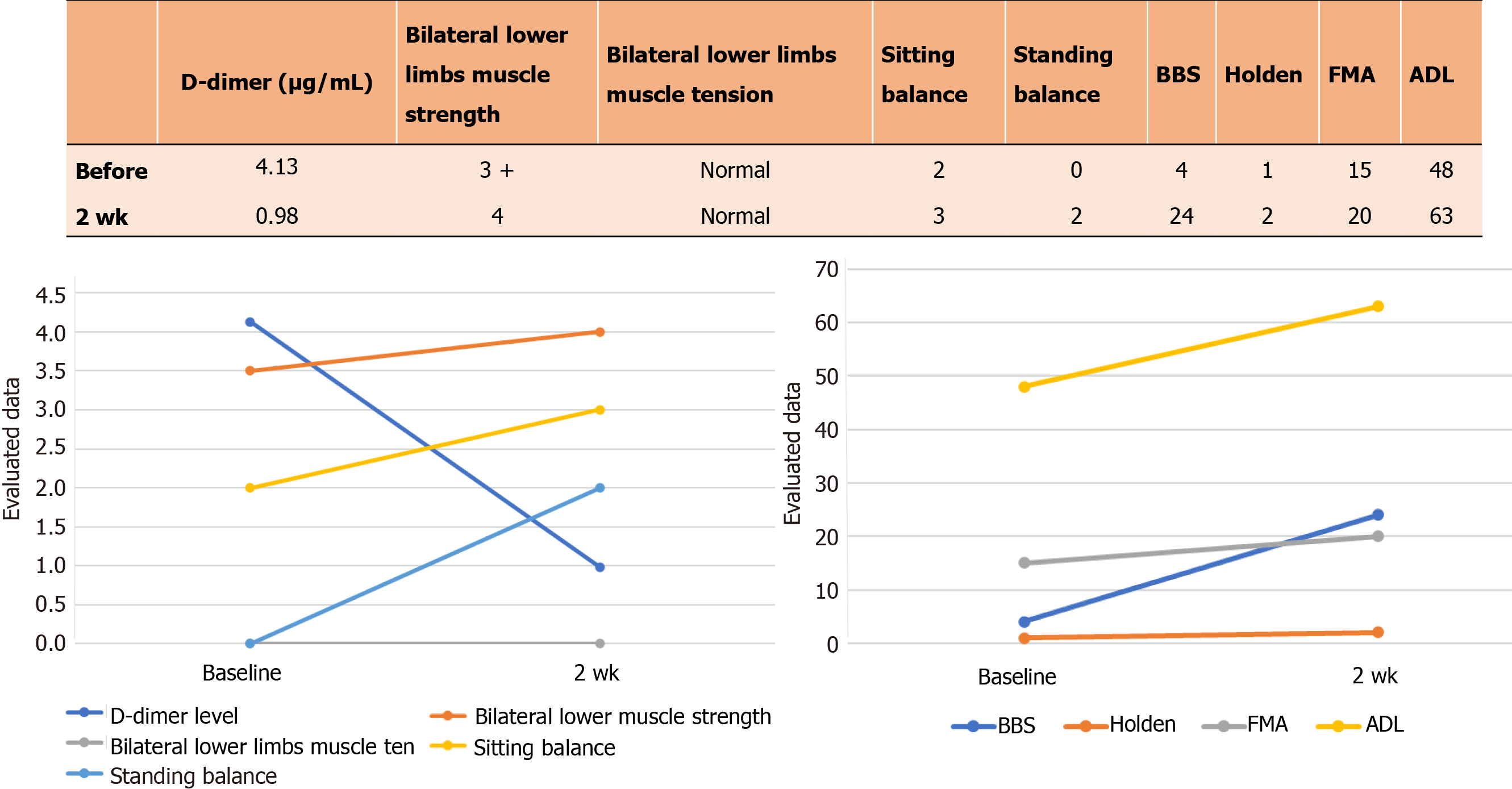
Figure 1 D-dimer and rehabilitation evaluation before and after 2 wk of treatment.
BBS: Berg Balance Scale; FMA: Fugl-Meyer assessment; ADL: Activities of daily living.
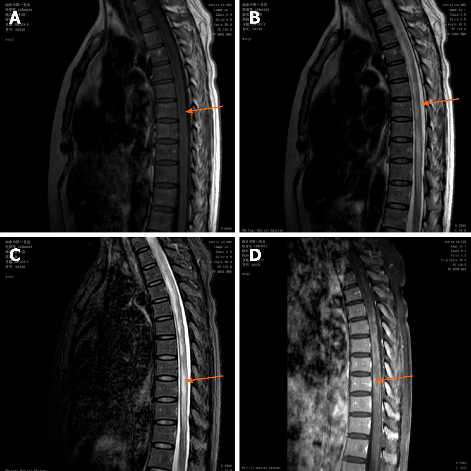
Figure 2 Thoracic and Lumbar magnetic resonance imaging on March 17, 2020 (illness day 7).
Strip-shaped abnormal signal shadow was observed in the medulla and the pathological changes in the spinal cord involved more than 3 spinal segments, which were represented by different signals in different phases of A, B, C, D. A: T1-weighted image (T1WI) was a low signal; B: T2-weighted image was a high signal; C: Short time inversion recovery (STIR) was a higher signal; D: T1WI-STIR enhancement was a higher signal and slight enhancement of the spinal cord margin.
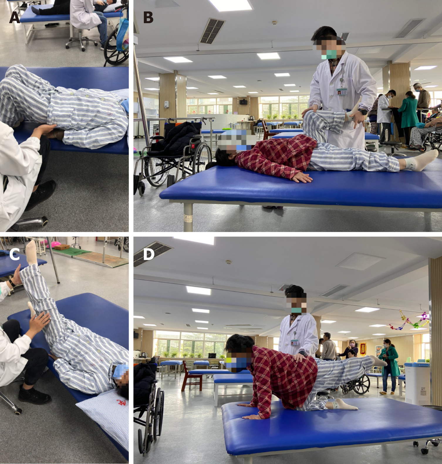
Figure 3 Rehabilitation treatment.
A: Active strength training of trunk muscles; B-D: Active strength training of lower limb muscles.
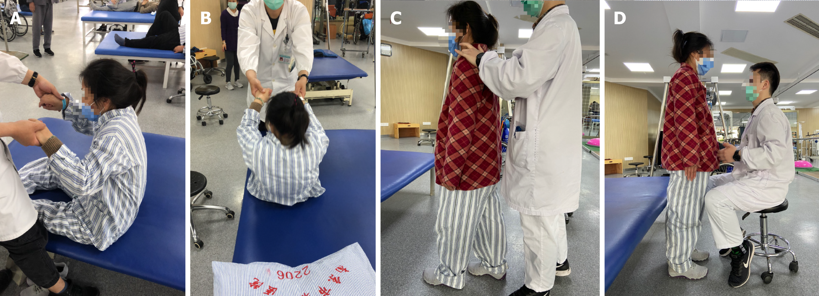
Figure 4 Rehabilitation treatment.
A and B: Balance training of sitting; C and D: Balance training of standing and walking.
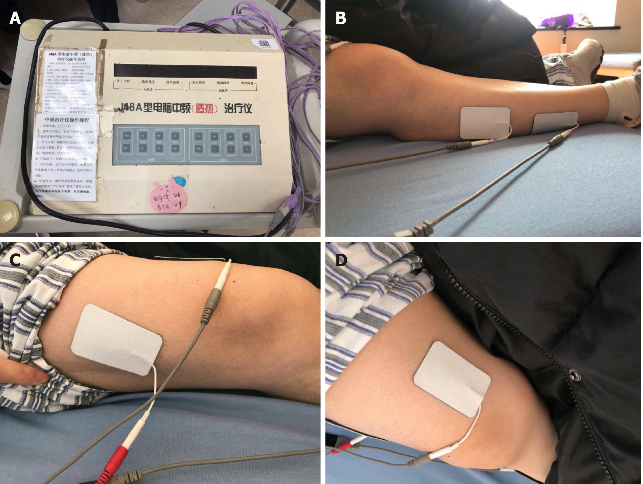
Figure 5 Neuromuscular electrical stimulation of lower limb muscles.
A: Computerized intermediate-frequency therapy apparatus; B: The electrodes were placed on the right calf; C and D: The electrodes were placed on the right thigh.
- Citation: Wang XJ, Xia P, Yang T, Cheng K, Chen AL, Li XP. Rehabilitation and pharmacotherapy of neuromyelitis optica spectrum disorder: A case report. World J Clin Cases 2021; 9(16): 3951-3959
- URL: https://www.wjgnet.com/2307-8960/full/v9/i16/3951.htm
- DOI: https://dx.doi.org/10.12998/wjcc.v9.i16.3951


