Copyright
©The Author(s) 2023.
World J Clin Cases. Apr 6, 2023; 11(10): 2246-2253
Published online Apr 6, 2023. doi: 10.12998/wjcc.v11.i10.2246
Published online Apr 6, 2023. doi: 10.12998/wjcc.v11.i10.2246
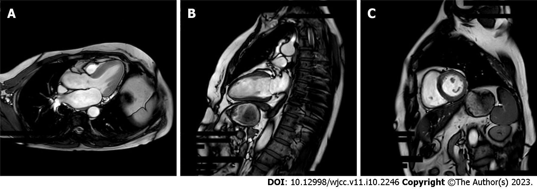
Figure 1 Magnetic resonance imaging results of a 56-year-old woman with hypertension, diabetes, chest tightness, and shortness of breath.
A: Functional imaging shows decreased left ventricular end-diastolic systole; B: The perfusion scan shows extensive subendocardial ischemia; C: The delayed scan shows partial myocardial fibrosis. Extensive myocardial ischemia was considered. Partial myocardial infarction causes abnormal cardiac function.
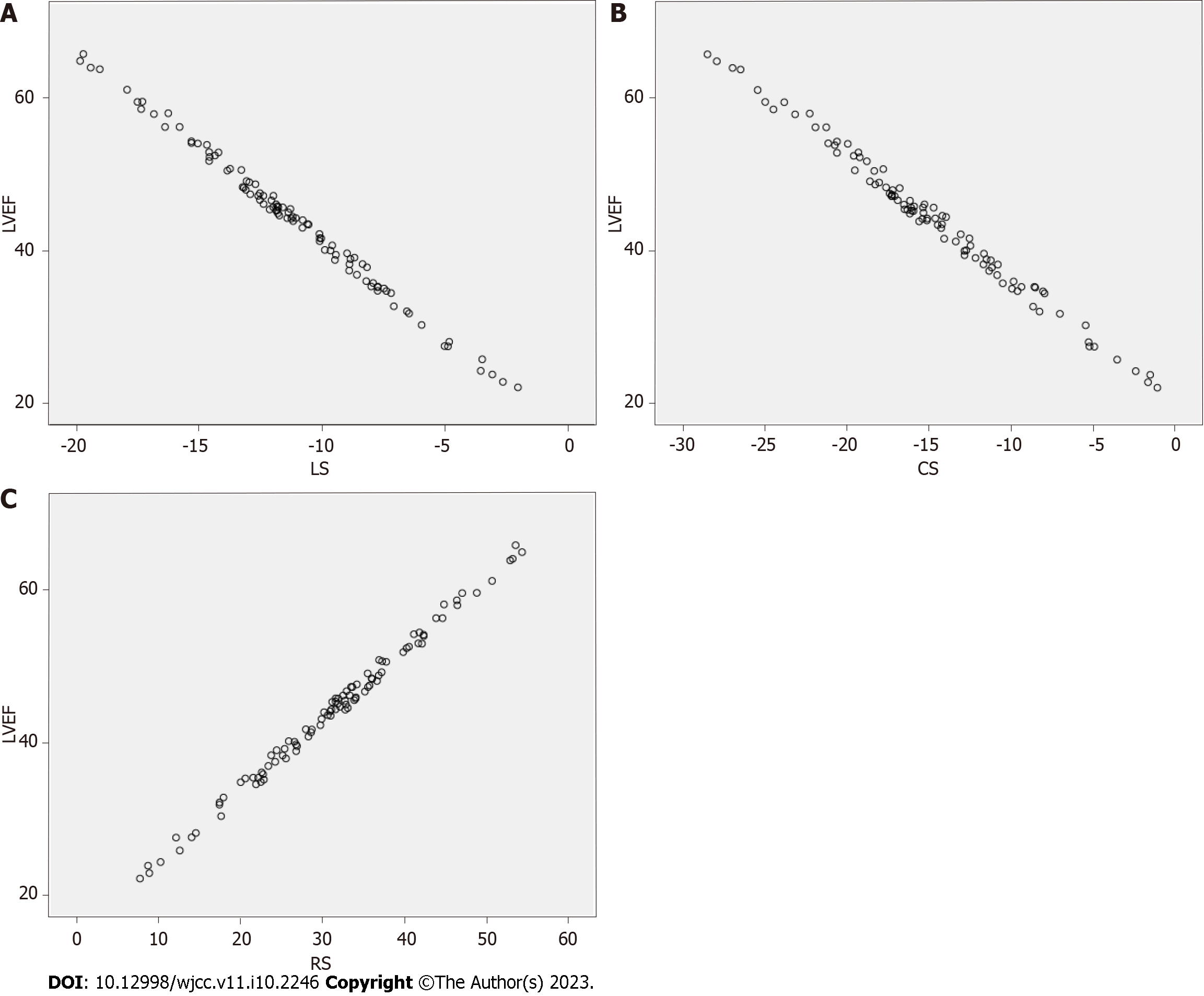
Figure 2 Interaction between the left ventricular ejection fraction and left ventricular strain.
A: Longitudinal strains; B: Circumferential strain; C: Radial strain. LVEF: Left ventricular ejection fraction; LS: Longitudinal strain; CS: Circumferential strain; RS: Radial strain.
- Citation: Gui HY, Liu SW, Zhu DF. Interaction between the left ventricular ejection fraction and left ventricular strain and its relationship with coronary stenosis. World J Clin Cases 2023; 11(10): 2246-2253
- URL: https://www.wjgnet.com/2307-8960/full/v11/i10/2246.htm
- DOI: https://dx.doi.org/10.12998/wjcc.v11.i10.2246


