Copyright
©The Author(s) 2022.
World J Clin Cases. Mar 16, 2022; 10(8): 2644-2649
Published online Mar 16, 2022. doi: 10.12998/wjcc.v10.i8.2644
Published online Mar 16, 2022. doi: 10.12998/wjcc.v10.i8.2644
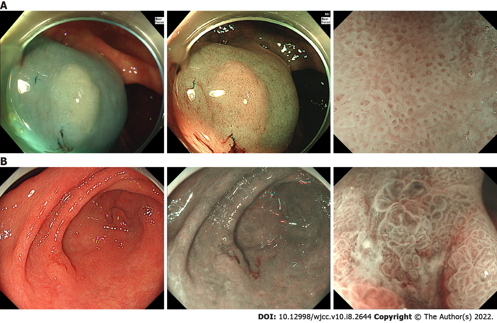
Figure 1 Narrow-band imaging magnified observation.
A: The first colonoscopy removed over 10 polyps and the diagnosis of serrated polyposis syndrome was established. A flat polyp with a size of 1.0 cm × 0.8 cm was observed in the ascending colon. The surface of the polyp was cloudy and the boundary was not clear. Type II open-shape pit pattern was seen by narrow-band imaging magnified observation after indigo carmine acetic acid staining; B: An elevated lesion was detected in the anterior wall of the gastric antrum at the gastroscopy. Upon white light endoscopy, a type IIc lesion approximately 1.2 cm × 1.0 cm in size could be seen in the anterior wall of the gastric antrum, with a small amount of white fur attached to the surface. Narrow-band imaging magnified observation showed the dividing line and the enlarged and irregular gland. No obvious abnormal blood vessels were found.
- Citation: Ning YZ, Liu GY, Rao XL, Ma YC, Rong L. Synchronized early gastric cancer occurred in a patient with serrated polyposis syndrome: A case report. World J Clin Cases 2022; 10(8): 2644-2649
- URL: https://www.wjgnet.com/2307-8960/full/v10/i8/2644.htm
- DOI: https://dx.doi.org/10.12998/wjcc.v10.i8.2644


