Copyright
©The Author(s) 2019.
World J Gastrointest Oncol. Jan 15, 2019; 11(1): 9-16
Published online Jan 15, 2019. doi: 10.4251/wjgo.v11.i1.9
Published online Jan 15, 2019. doi: 10.4251/wjgo.v11.i1.9
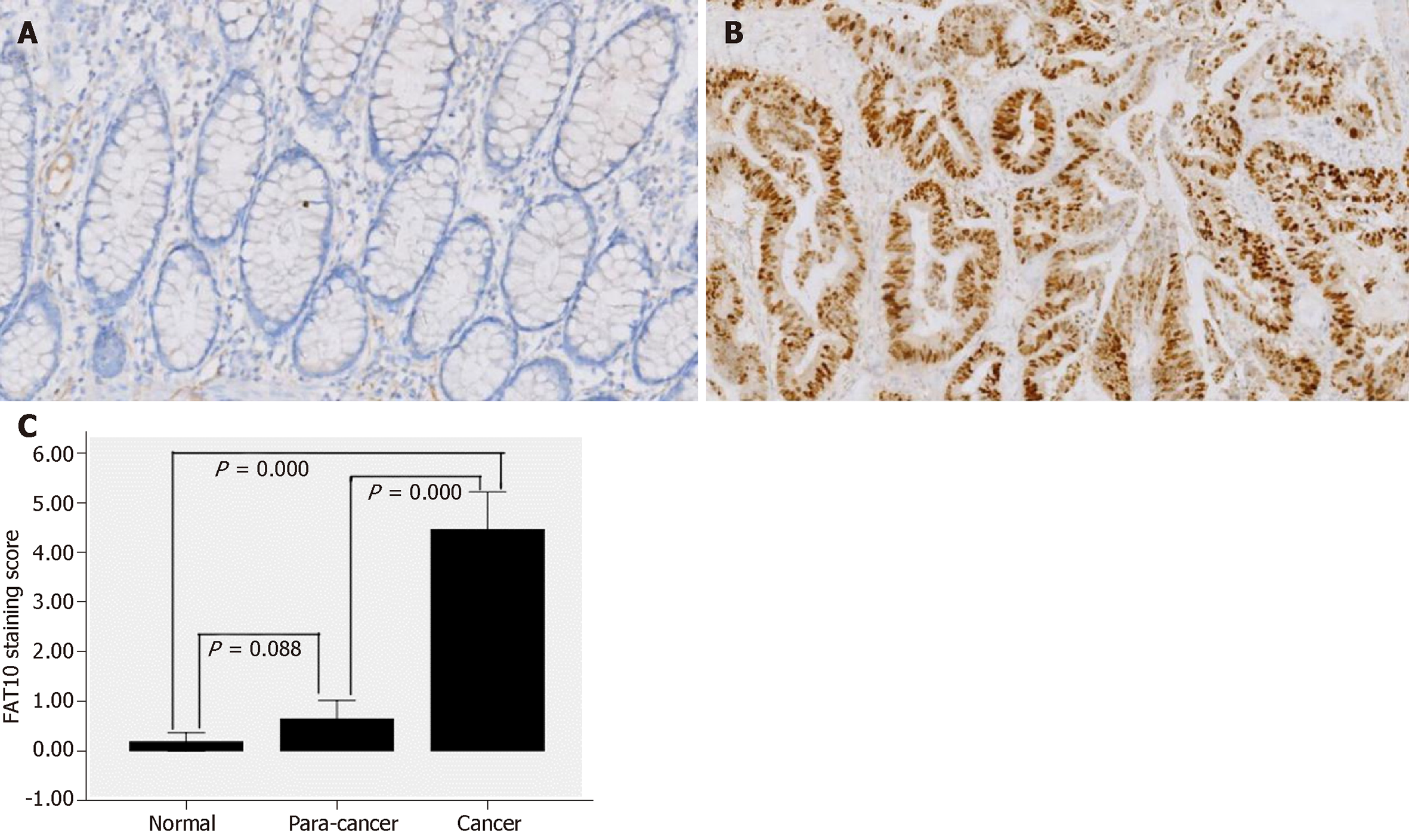
Figure 1 Immunohistochemical staining for human leukocyte antigen F-associated transcript 10 in colorectal tissues (original magnification, 100×).
A: Human leukocyte antigen F-associated transcript 10 (FAT10) expression is negative in para-cancer tissue; B: FAT10 expression is strongly positive in moderately differentiated colorectal adenocarcinoma; C: Semi-quantitative analysis of FAT10 expression in normal colorectal mucosal tissue, para-cancer tissue, and colorectal cancer tissue.
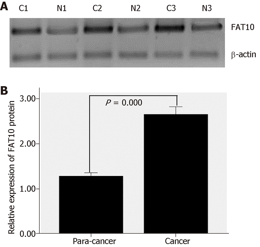
Figure 2 Western blot analysis of human leukocyte antigen F-associated transcript 10 expression in colorectal cancer and para-cancer tissues.
A: Western blot analysis; B: Relative expression. FAT10: Human leukocyte antigen F-associated transcript 10.
- Citation: Zhang CY, Sun J, Wang X, Wang CF, Zeng XD. Clinicopathological significance of human leukocyte antigen F-associated transcript 10 expression in colorectal cancer. World J Gastrointest Oncol 2019; 11(1): 9-16
- URL: https://www.wjgnet.com/1948-5204/full/v11/i1/9.htm
- DOI: https://dx.doi.org/10.4251/wjgo.v11.i1.9


