Copyright
©The Author(s) 2021.
World J Clin Cases. Aug 6, 2021; 9(22): 6544-6551
Published online Aug 6, 2021. doi: 10.12998/wjcc.v9.i22.6544
Published online Aug 6, 2021. doi: 10.12998/wjcc.v9.i22.6544
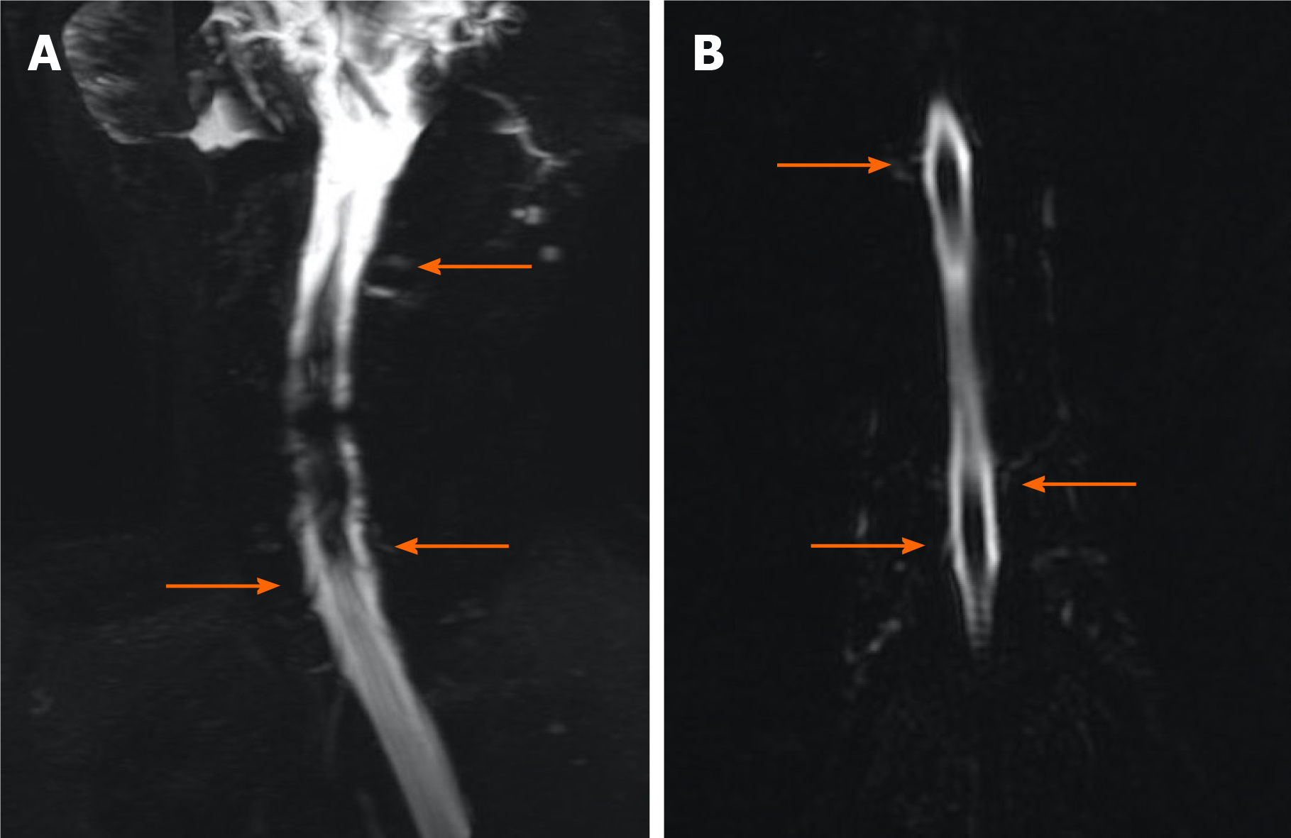
Figure 2 Magnetic resonance myelography imaging of the spine performed before epidural blood patch therapy.
A: Magnetic resonance myelography (MRM) shows multiple sites of cerebrospinal fluid (CSF) leakage at cervicothoracic junctions on day 9 (orange arrows); B: MRM shows multiple sites of CSF leakage in the thoracic region on day 9 (orange arrows).
- Citation: Wei TT, Huang H, Chen G, He FF. Management of an intracranial hypotension patient with diplopia as the primary symptom: A case report . World J Clin Cases 2021; 9(22): 6544-6551
- URL: https://www.wjgnet.com/2307-8960/full/v9/i22/6544.htm
- DOI: https://dx.doi.org/10.12998/wjcc.v9.i22.6544


