Published online Jul 16, 2021. doi: 10.12998/wjcc.v9.i20.5605
Peer-review started: February 9, 2021
First decision: March 25, 2021
Revised: April 1, 2021
Accepted: May 15, 2021
Article in press: May 15, 2021
Published online: July 16, 2021
Processing time: 147 Days and 20 Hours
Superficial CD34-positive fibroblast tumors (SCPFTs) are newly recognized fibroblast and myofibroblast tumors representing intermediate tumors. To the best of our knowledge, fewer than 50 cases have been reported. Perianal SCPFT has not been previously reported.
A 55-year-old man was hospitalized upon discovering a painless perianal lump 10 d prior. Physical examination showed a lump of approximately 3 cm × 4 cm in the 7 to 8 o’clock direction in the perianal area. Perianal abscess was considered the primary diagnosis. Lump removal surgery was performed under epidural anesthesia. Postoperative pathology showed a well-circumscribed, soft tissue-derived, spindle-cell tumor with strong CD34 positivity by immunohistochemistry. The final diagnosis was perianal SCPFT. There were no complications, and the patient was followed for more than 8 mo without recurrence or metastasis.
We report a case of perianal superficial CD34-positive fibroblast tumor. This rare mesenchymal neoplasm has distinctive histomorphology, which is important for diagnosis. Comprehensive consideration of clinical information, imaging, histology, and immunohistochemistry is important for diagnosis.
Core Tip: We present a new case of perianal superficial CD34-positive fibroblast tumor. Surgery is the main treatment for superficial painless slowly growing masses. Postoperative immunohistochemical examination showed that strong positivity for CD34 and good prognosis were the characteristics of the case.
- Citation: Long CY, Wang TL. Perianal superficial CD34-positive fibroblastic tumor: A case report. World J Clin Cases 2021; 9(20): 5605-5610
- URL: https://www.wjgnet.com/2307-8960/full/v9/i20/5605.htm
- DOI: https://dx.doi.org/10.12998/wjcc.v9.i20.5605
Superficial CD34-positive fibroblast tumors (SCPFTs) are newly recognized fibroblast and myofibroblast tumors representing intermediate tumors. SCPFT was first reported in 2014[1]. To date, less than 50 cases have been reported. Perianal SCPFT has not been previously reported[2]. Here, we report a case that was misdiagnosed as a perianal abscess before surgery. Informed consent for the publication of these data was obtained from the patient.
A 55-year-old man was hospitalized after he discovered a painless perianal mass.
The patient’s symptoms started 10 d prior to presentation.
The patient had no relevant previous medical history.
The patient’s family history was unremarkable.
A lump approximately 3 cm × 4 cm could be felt in the 7 to 8 o’clock direction of the perianal area.
After admission to the inpatient ward, laboratory examinations were carried out, which included routine blood tests (Table 1), routine tests for stool plus occult blood, and tests for liver and kidney function, electrolytes, blood coagulation function, and tumor biomarkers. Preoperative examinations ruled out hepatitis B, hepatitis C, syphilis, and human immunodeficiency virus. All results were within normal ranges.
| Inflammatory factor | Tumor biomark | ||
| White blood cell count | 7.55 × 10⁹/L | AFP | 4.08 ng/mL |
| Neutrophil count | 4.28 × 10⁹/L | CA19-9 | 11.51 U/mL |
| Neutrophil percentage | 56.6% | CA125 | 7.6 U/mL |
| High-sensitivity C-reactive protein | 0.46 mg/L | CEA | 1.97 ng/mL |
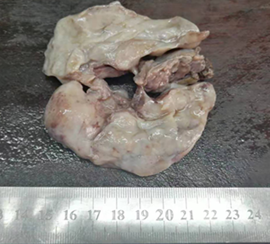
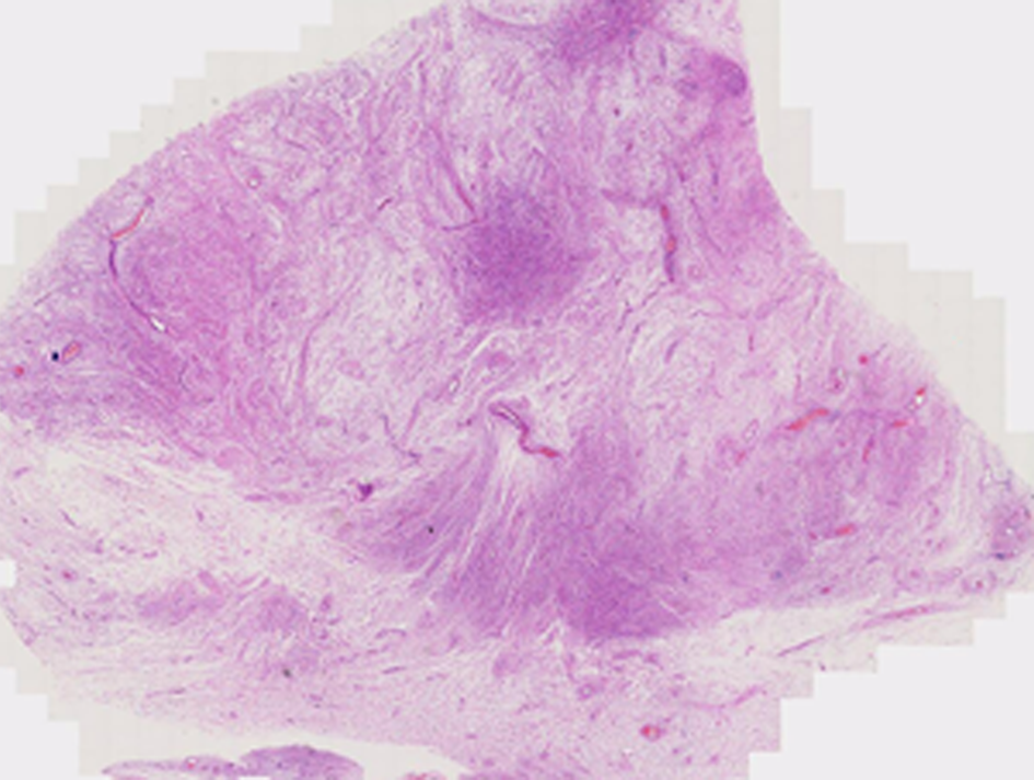
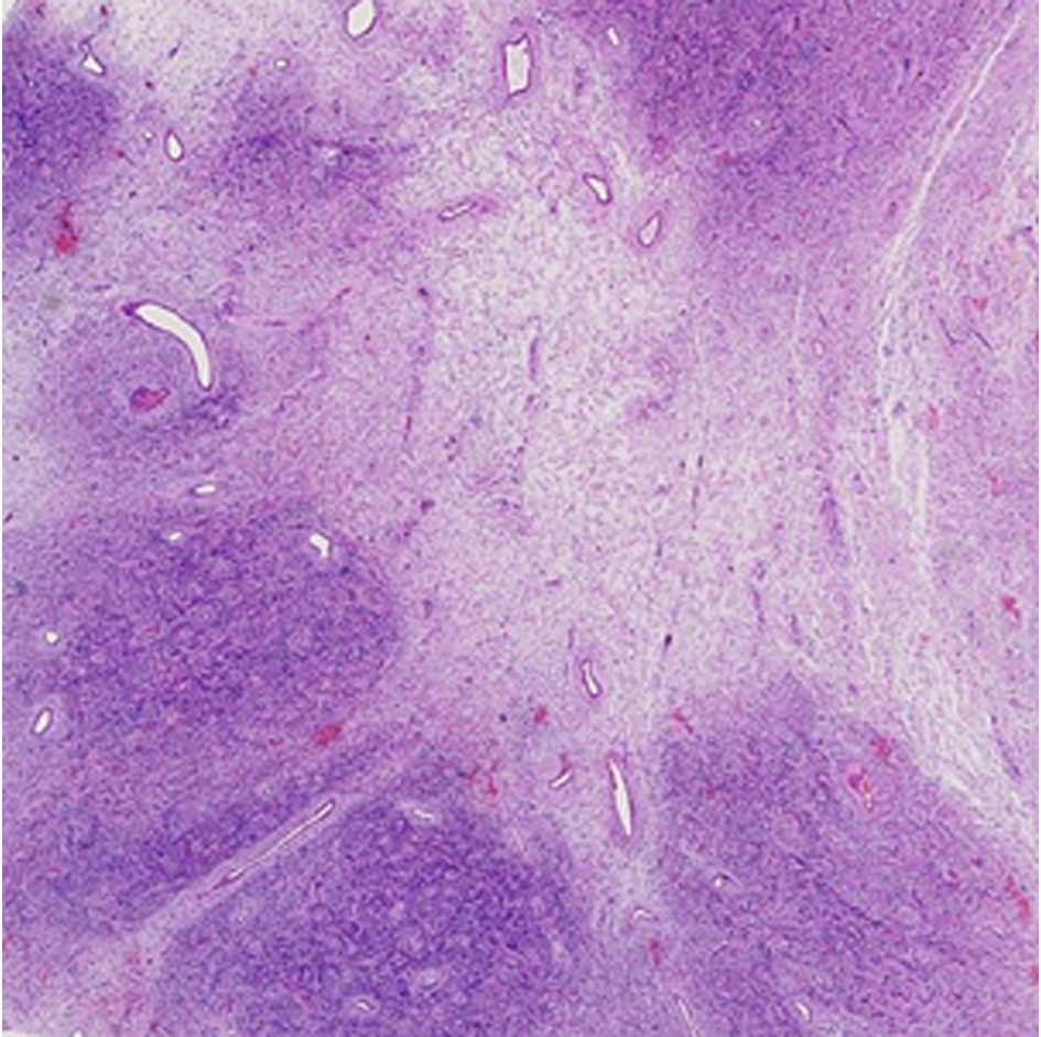
Postoperative pathology showed that a lump approximately 8 cm × 6.5 cm × 5 cm with a clear boundary, regional capsule, surface color of gray or taupe, interior color of gray, likely nodules, and mucoid changes in some areas was observed (Figures 1-3).
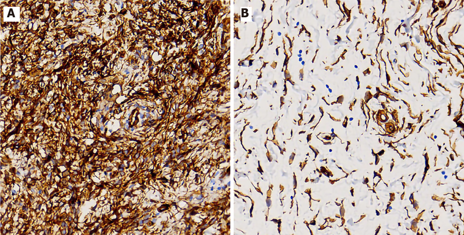
Immunohistochemistry showed that the tumor cells were diffusely and strongly positive for CD34 and vimentin, but negative for CD31, S100, desmin, EMA, SMA, CD117, Dog-1, CK-P, INI1, CD68, CD99, STAT6, β-catenin, HMB45, and ALK (D5F3) (Figure 4). The Ki-67 index was < 1%.
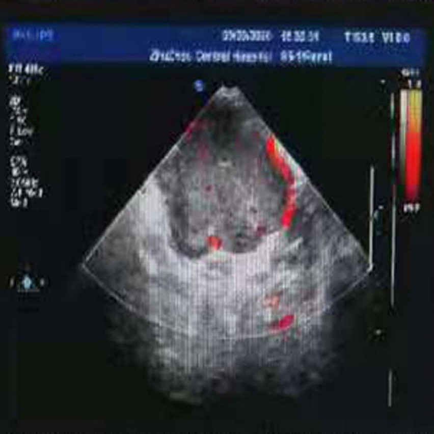
Ultrasound showed a 7.9 cm × 7.6 cm cystic mass in the 1 to 5 o’clock direction in the knee-chest position. The border was clear with poor entrant sound and rear echo enhancement. Many vascular signals could be detected around the mass (Figure 5).
SCPFT.
Lump removal surgery was performed under epidural anesthesia.
There were no complications, and the patient was followed for more than 8 mo without recurrence or metastasis.
SCPFTs are mostly slow-growing, painless lumps, occurring in patients with a median age of 35 years (age range, 20-76 years) with a slight male preponderance[3-8]. It most commonly occurs in the lower limb, thigh, buttock, shoulder, and upper arm. The location in the perianal region was not previously reported. Our patient had a small red mass but had no fever before surgery and no fever or pain, and routine blood examination was normal. It was misdiagnosed as a perianal abscess due to the unusual disease location combined with B ultrasound results. Perianal abscess often manifests as an inflammatory mass with obvious pain. The total number of leukocytes and proportion of neutrophils can be increased on routine blood examinations. CD34 expression status on immunohistochemistry is the most important discriminatory factor.
Histologically, SCPFT can vary and has many forms without unique histological morphological characteristics, and the disease can be easily misdiagnosed as other mesenchymal tumors. The features of SCPFT include the following: (1) It is a slow-growing, painless lump; (2) The tumor is confined to the deep dermis or superficial fibroadipose tissue; (3) Tumor cells are composed of plump spindle to epithelioid cells[9]; and (4) CD34 is strongly positive on immunohistochemistry, with partial cellular expression of keratin, no INI1 expression, and a low Ki67 proliferative index[10].
To date, surgical resection has been used to treat SCPFT. Only one patient had lymph node metastasis after the operation[3]. No recurrence or metastasis was reported.
This is the first reported case of perianal SCPFT. Due to the novelty of this tumor, the long-term prognosis is not clear. Therefore, it is necessary to accumulate more cases and conduct long-term follow-up.
Manuscript source: Unsolicited manuscript
Specialty type: Surgery
Country/Territory of origin: China
Peer-review report’s scientific quality classification
Grade A (Excellent): 0
Grade B (Very good): B
Grade C (Good): C, C
Grade D (Fair): 0
Grade E (Poor): 0
P-Reviewer: Barosi G, Enomoto H S-Editor: Yan JP L-Editor: Wang TQ P-Editor: Li JH
| 1. | Carter JM, Weiss SW, Linos K, DiCaudo DJ, Folpe AL. Superficial CD34-positive fibroblastic tumor: report of 18 cases of a distinctive low-grade mesenchymal neoplasm of intermediate (borderline) malignancy. Mod Pathol. 2014;27:294-302. [RCA] [PubMed] [DOI] [Full Text] [Cited by in Crossref: 58] [Cited by in RCA: 71] [Article Influence: 6.5] [Reference Citation Analysis (1)] |
| 2. | Lin TL, Yang CS, Juan CK, Weng YC, Chen YJ. Superficial CD34-Positive Fibroblastic Tumor: A Case Report and Review of the Literature. Am J Dermatopathol. 2020;42:68-71. [RCA] [PubMed] [DOI] [Full Text] [Cited by in Crossref: 4] [Cited by in RCA: 4] [Article Influence: 0.8] [Reference Citation Analysis (0)] |
| 3. | Lao IW, Yu L, Wang J. Superficial CD34-positive fibroblastic tumour: a clinicopathological and immunohistochemical study of an additional series. Histopathology. 2017;70:394-401. [RCA] [PubMed] [DOI] [Full Text] [Cited by in Crossref: 25] [Cited by in RCA: 38] [Article Influence: 4.2] [Reference Citation Analysis (0)] |
| 4. | Hendry SA, Wong DD, Papadimitriou J, Robbins P, Wood BA. Superficial CD34-positive fibroblastic tumour: report of two new cases. Pathology. 2015;47:479-482. [RCA] [PubMed] [DOI] [Full Text] [Cited by in Crossref: 17] [Cited by in RCA: 19] [Article Influence: 2.1] [Reference Citation Analysis (0)] |
| 5. | Wada N, Ito T, Uchi H, Nakahara T, Tsuji G, Yamada Y, Oda Y, Furue M. Superficial CD34-positive fibroblastic tumor: A new case from Japan. J Dermatol. 2016;43:934-936. [RCA] [PubMed] [DOI] [Full Text] [Cited by in Crossref: 18] [Cited by in RCA: 19] [Article Influence: 2.1] [Reference Citation Analysis (0)] |
| 6. | Li W, Molnar SL, Mott M, White E, De Las Casas LE. Superficial CD34-positive fibroblastic tumor: Cytologic features, tissue correlation, ancillary studies, and differential diagnosis of a recently described soft tissue neoplasm. Diagn Cytopathol. 2016;44:926-930. [RCA] [PubMed] [DOI] [Full Text] [Cited by in Crossref: 11] [Cited by in RCA: 11] [Article Influence: 1.2] [Reference Citation Analysis (0)] |
| 7. | Donaldson MR, Weber LA. Superficial CD34-Positive Fibroblastic Tumor Treated With Mohs Micrographic Surgery. Dermatol Surg. 2017;43:1489-1491. [RCA] [PubMed] [DOI] [Full Text] [Cited by in Crossref: 5] [Cited by in RCA: 5] [Article Influence: 0.7] [Reference Citation Analysis (0)] |
| 8. | Sood N, Khandelia BK. Superficial CD34-positive fibroblastic tumor: A new entity; case report and review of literature. Indian J Pathol Microbiol. 2017;60:377-380. [RCA] [PubMed] [DOI] [Full Text] [Cited by in Crossref: 5] [Cited by in RCA: 6] [Article Influence: 0.9] [Reference Citation Analysis (0)] |
| 9. | Zemheri E, Karadag AS, Yılmaz İ. Superficial CD34-Positive Fibroblastic Tumor: Report of an Extremely Rare Entity. Indian J Dermatol. 2020;65:526-529. [RCA] [PubMed] [DOI] [Full Text] [Cited by in Crossref: 2] [Cited by in RCA: 2] [Article Influence: 0.4] [Reference Citation Analysis (0)] |
| 10. | Yu L, Wang J. [Updates on pathology of soft tissue tumors]. Zhonghua Bing Li Xue Za Zhi. 2013;42:145-146. [RCA] [PubMed] [DOI] [Full Text] [Cited by in RCA: 1] [Reference Citation Analysis (0)] |


