Copyright
©The Author(s) 2020.
World J Clin Cases. Nov 26, 2020; 8(22): 5684-5689
Published online Nov 26, 2020. doi: 10.12998/wjcc.v8.i22.5684
Published online Nov 26, 2020. doi: 10.12998/wjcc.v8.i22.5684
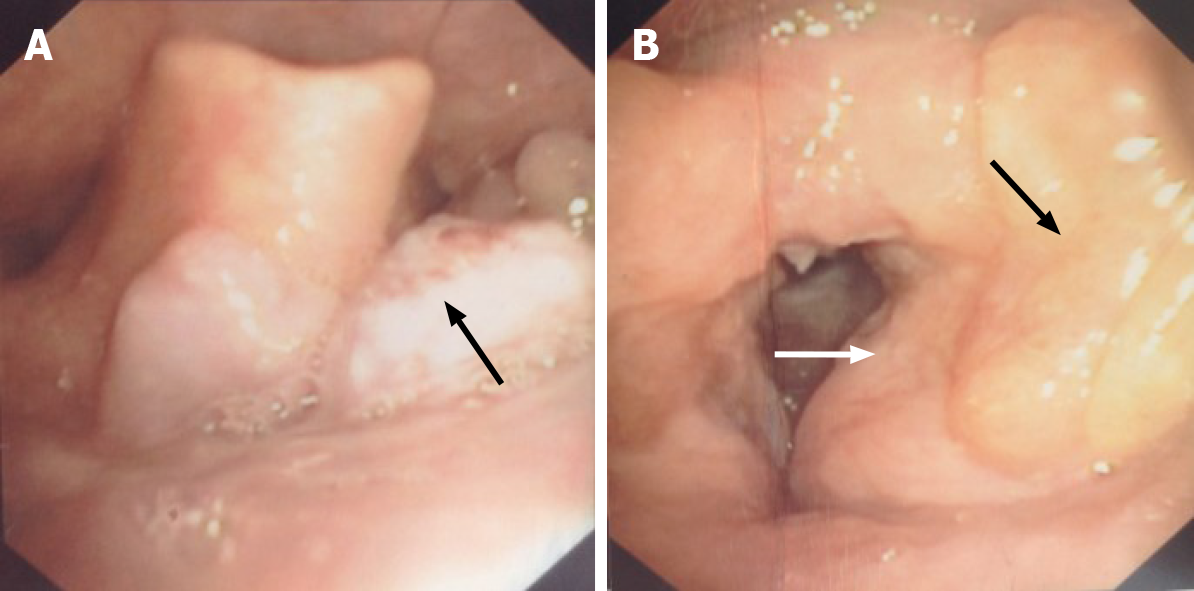
Figure 1 Fiber laryngoscopy findings before surgery.
A: Fiber laryngoscopy showed a neoplasm with a coarse surface (black arrow) near the tongue base; B: Left vocal cord was covered by swelling and thickening ventricular fold (white arrow), and yellow sediment was observed near the left ventricular fold (black arrow). No lesion was found on the right vocal cord.
- Citation: Song X, Yang J, Lai Y, Zhou J, Wang J, Sun X, Wang D. Localized amyloidosis affecting the lacrimal sac managed by endoscopic surgery: A case report. World J Clin Cases 2020; 8(22): 5684-5689
- URL: https://www.wjgnet.com/2307-8960/full/v8/i22/5684.htm
- DOI: https://dx.doi.org/10.12998/wjcc.v8.i22.5684


