Published online Jul 16, 2024. doi: 10.12998/wjcc.v12.i20.4446
Revised: May 15, 2024
Accepted: May 29, 2024
Published online: July 16, 2024
Processing time: 81 Days and 16.1 Hours
Diabetic foot ulcers are caused by a variety of factors, including peripheral neu
The purpose of this case report is to highlight the efficacy of polydeoxyribonucleotide (PDRN) injection as a non-surgical treatment option for diabetic foot ulcers on the dorsum of the foot, particularly in patients who choose against surgical intervention. This case report presents two cases of diabetic foot ulcers located on the dorsum of the foot that were successfully treated with PDRN injection as a non-surgical intervention.
If the patient declines surgery for diabetic ulcers with Wagner grade II or below, PDRN injection can be effective if necrotic tissue is removed and the wound bed kept clean.
Core Tip: If a diabetic foot wound of Wager grade II or less is located on the dorsum of the foot and is not amenable to surgical treatment, polydeoxyribonucleotide injection combined with a minimally invasive treatment of debridement under local anesthesia may help wound healing in patients who are not amenable to surgical treatment.
- Citation: Ha Y, Kim JH, Kim J, Kwon H. Non-surgical treatment of diabetic foot ulcers on the dorsum of the foot with polydeoxyribonucleotide injection: Two case reports. World J Clin Cases 2024; 12(20): 4446-4451
- URL: https://www.wjgnet.com/2307-8960/full/v12/i20/4446.htm
- DOI: https://dx.doi.org/10.12998/wjcc.v12.i20.4446
Diabetic foot ulcers are caused by a variety of factors, including peripheral neuropathy, peripheral arterial disease, impaired wound healing mechanisms, and repetitive trauma, and are a fatal condition that affects 19%-35% of people with diabetes mellitus and can lead to amputation and death[1]. Proper care of diabetic foot ulcers is an important factor to increase patient outcome and favorable prognosis[2]. Patients with diabetic foot ulcer on the dorsum of the foot have often been treated surgically using techniques like free flap[3] or local flaps[4]. However, the right non-surgical therapy must be chosen if surgical choices are contraindicated or if the patient prefers conservative treatment over surgery.
It is important to choose an effective non-surgical treatment option if the patient declines surgical reconstruction because foot ulcers in the midfoot heal more slowly and require longer than ulcers in the toe[5]. Additionally, if possible, amputation should be performed distally[6], but ulcers in the dorsum of the foot may require a more proximal ampu
Free flap surgery, also known as free tissue transfer, involves transferring tissue with its blood supply from one part of the body to another for reconstructive purposes. The procedure allows for the repair of diabetic foot ulcers by using healthy tissue from elsewhere in the body. Through microsurgery techniques, the transferred tissue is reconnected to blood vessels at the recipient site. Free flap is the preferred surgical treatment for diabetic foot ulcers with good prognosis and increased survival rate[7]. However, free flap reconstruction requires careful surgical planning, a long stay in the hospital, and can be accompanied by potential surgical complications[8] such as flap failure, donor site morbidity, infection, or wound dehiscence, as well as a shallow learning curve for the surgeon to become proficient in microsurgery.
A substance generated from salmon DNA called PDRN has shown extraordinary abilities in the processes of tissue regeneration, wound healing, and anti-inflammatory responses. Many in vitro studies[9,10], animal studies[11,12], and clinical studies[13] have demonstrated its effectiveness, and it is expanding its scope by demonstrating its effectiveness in the reconstructive field.
The purpose of this case report is to highlight the efficacy of PDRN injection as a non-surgical treatment option for diabetic foot ulcers on the dorsum of the foot, particularly in patients who choose against surgical intervention. This case report presents two cases of diabetic foot ulcers located on the dorsum of the foot, successfully treated with PDRN injection as a non-surgical intervention. Both cases involved patients with longstanding diabetes and chronic non-healing ulcers, warranting alternative therapeutic options due to the risks associated with surgical procedures and the limited response to conventional wound care measures.
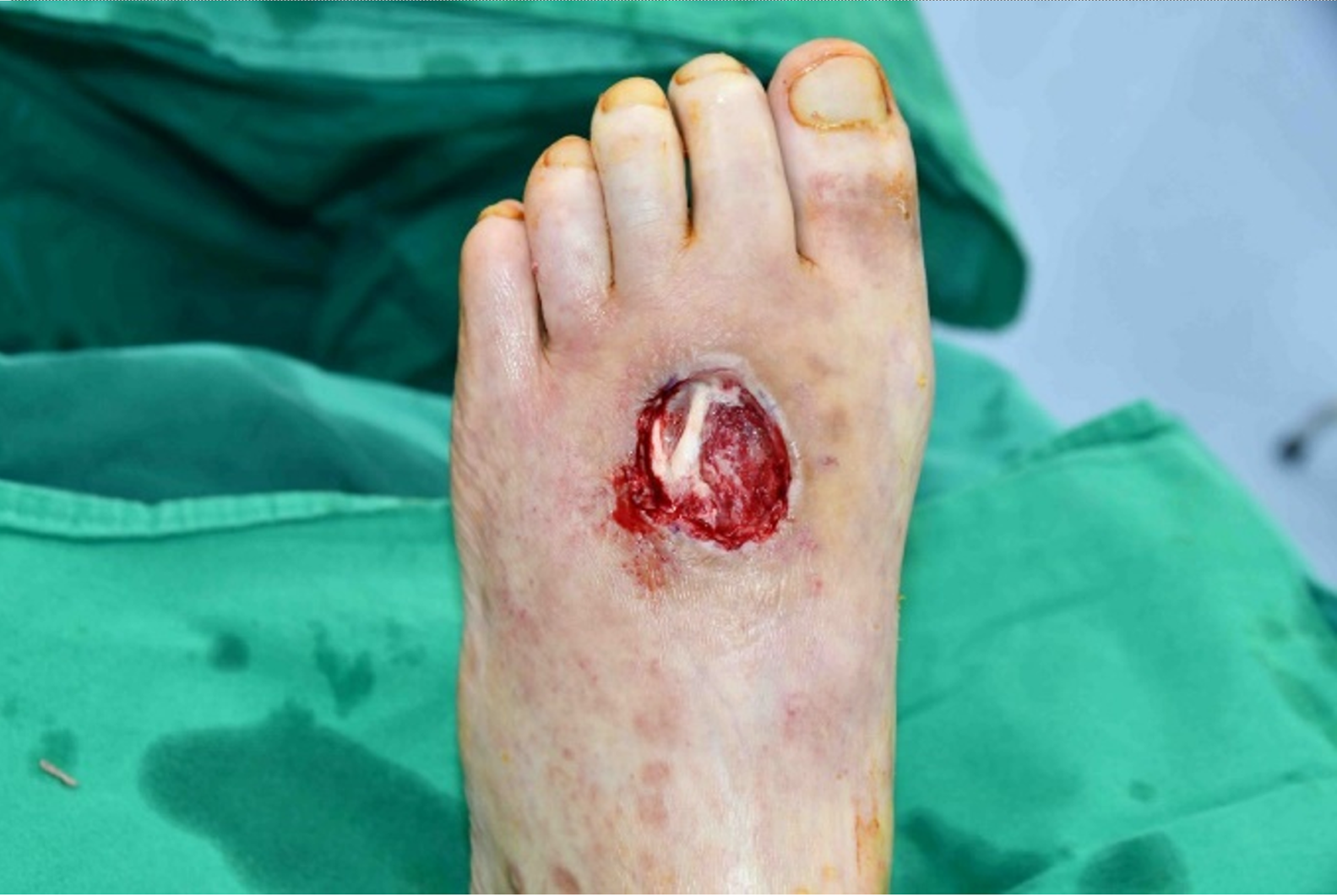
Case 1: A 68-year-old man who had a soft tissue defect to the left dorsum of his foot (Wagner grade II) measuring about 3 cm 3 cm and present for 6 mo came to the plastic surgery outpatient department (Figure 1).
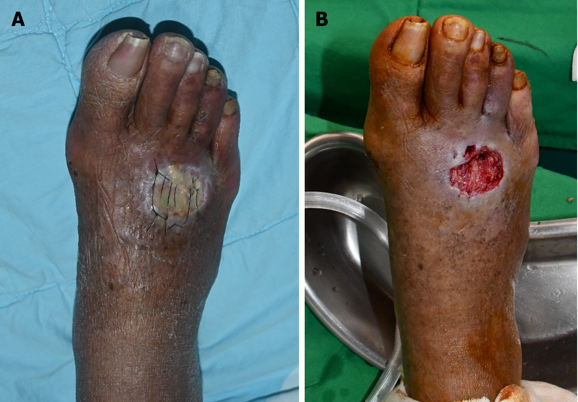
Case 2: A 70-year-old woman who had a soft tissue defect to the right dorsum of her foot (Wagner grade II) measuring about 2 cm 2.5 cm and present for 3 mo came to the plastic surgery outpatient department (Figure 2).
Case 1: The patient received conservative care with regular dressings, but the wound condition did not improve.
Case 2: The patient first had a small wound that another hospital tried to stitch closed. However, her wound did not heal, and she eventually came to the plastic surgery outpatient department with wet eschar and significant necrosis.
Case 1: This patient did not have a family history of diabetic foot ulcers.
Case 2: This patient did not have a family history of diabetic foot ulcers.
Case 1: His ankle-brachial index (ABI) was 0.96.
Case 2: Her ABI was 0.91.
Case 1: His HbA1c was 9.6%, indicating poor blood sugar control.
Case 2: Her HbA1c was 8.8%, suggesting poor blood sugar control.
No additional radiologic study was performed.
Hard-to-heal Diabetic foot ulcers.
Case 1: We first advised surgery, such as a free flap, but the patient declined because of anxiety over potential complications. In consideration of this, we began conservative treatment with PDRN and adequate glycemic control. PDRN (5.625 mg) was intralesionally injected into the subcutaneous layer of the wound once a week for a total of 8 wk after we debrided the necrotic tissue of the wound at the patient's initial visit. The patient applied a wet-to-dry gauze dressing to the wound daily while receiving therapy.
Case 2: As in case 1, we first recommended surgery, but the patient refused. PDRN (5.625 mg) was injected in a perilesional manner into the subcutaneous layer once a week after the necrotic tissues had been surgically debrided, and daily wet-to-dry gauze dressing was applied as in case 1.
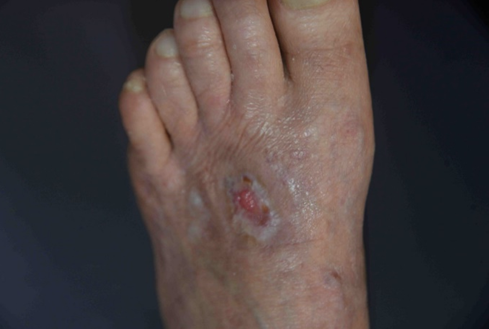
Case 1: After about 2 wk of dressing and two PDRN treatments, there was no noticeable healing effect, so we debrided the unhealthy tendon (extensor digitorum brevis) that was exposed during the dressing, and the wound began to show noticeable wound healing.
Eight weeks later, the patient's diabetic foot ulcer had epithelialized and had fully recovered without any sequelae (Figure 3). He recovered from his diabetic foot ulcer, and it did not get worse over the following 2 mo.
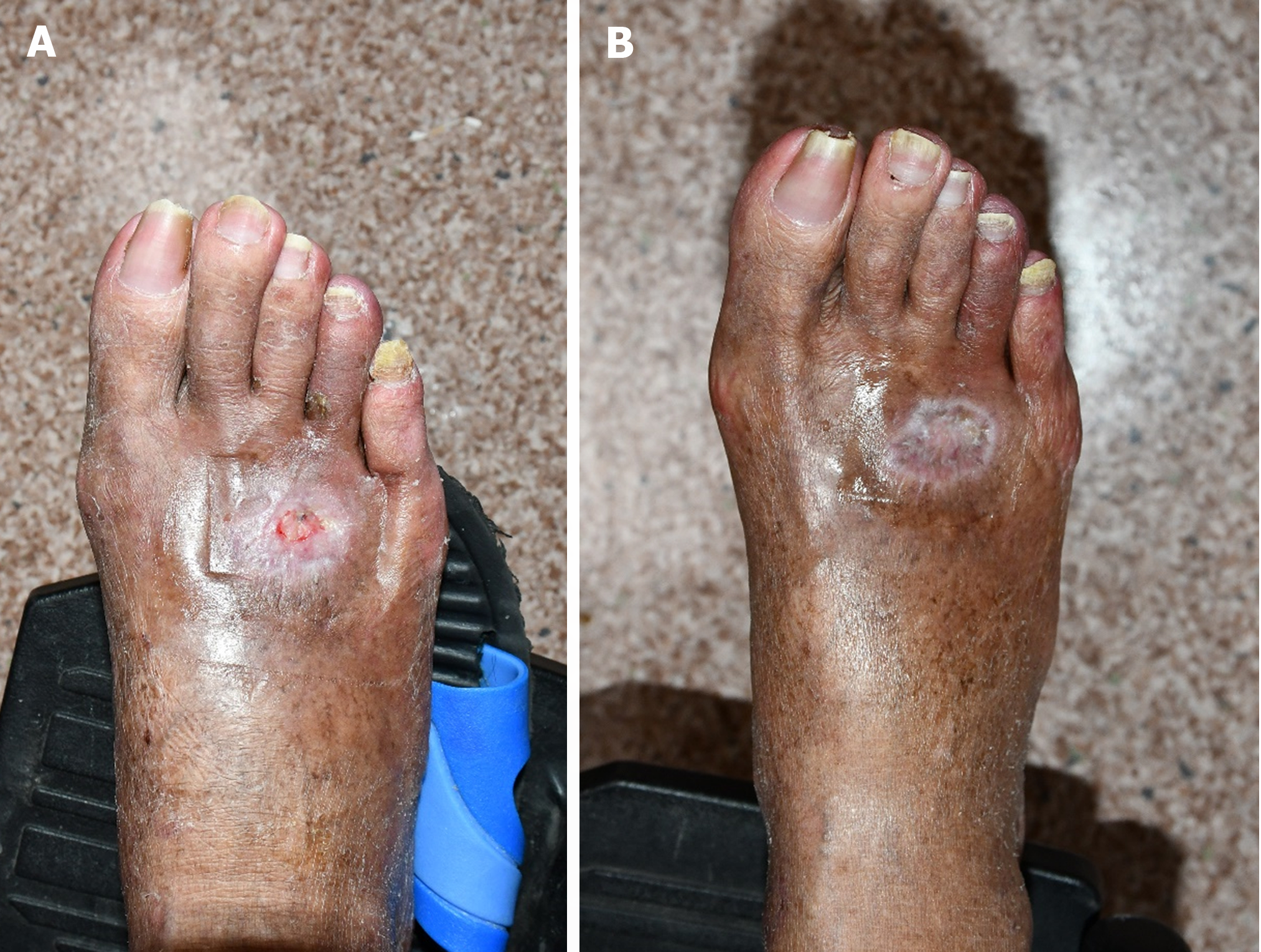
Case 2: After 8 wk, the diabetic foot ulcer on the dorsum of her foot had healed and left no significant sequelae (Figure 4). There were no complications at the 1-mo follow-up.
If surgical repair for a diabetic foot ulcer on the dorsum of the foot is available, it is the usual treatment choice. However, there are few conservative treatment options available for the patients who choose to refuse surgical procedures. Collagen dressing[14], acellular fish skin dressing[15], negative-pressure wound therapy[16], and other treatments are known to aid in the healing of diabetic foot ulcers, but their broad usage is limited due to the high cost and difficulties in carrying the device. However, PDRN had a positive effect on wound healing in these cases with just one weekly injection and without the disadvantages described above. Therefore, it can be another treatment option for patients who refuse surgical treatment.
The observed healing response in the dorsum of the foot following PDRN injections supports prior studies[9-12] that demonstrated PDRN's therapeutic potential in stimulating tissue regeneration. PDRN's angiogenic features are thought to play a critical role in encouraging blood vessel creation and increasing local microcirculation, resulting in improved wound healing.
In diabetic foot ulcer patients with severe vascular compromise, revascularization is a critical intervention, and we believe that conservative treatment with PDRN alone may not be appropriate in patients with an ABI of 0.9 or less.
In previous clinical studies applying PDRN, PDRN was applied almost daily (5 d a week) by various routes (perilesional or intramuscular)[17,18], but it is difficult to apply it 5 d a week in reality due to economic problems, but in this case, we demonstrate that only once a week perilesional injection is effective.
The dorsal surface of the foot is characterized by the presence of tendons just below the thin soft tissue, and wounds exposing the tendons are difficult to reconstruct with relatively minor surgery such as split-thickness skin graft (STSG) due to poor vascularity of the tendons[19]. In addition, due to the poor vascularity of the tendon, tissue granulation and epithelialization are difficult, making reconstruction difficult even with conservative treatment. As observed in Case 1 above, if the tendon is exposed, sacrifice of the tendon may be considered if damaged, non- functional and unlikely to result in significant comorbidity.
When performing STSG reconstruction on a wound with tendon exposure, it has been reported that flipping the tendon over allows for better STSG uptake due to the presence of vascularized connective tissue[19]. Therefore, if it is difficult to sacrifice the tendon, it is possible to consider applying PDRN after flipping the tendon to expose the vascularized connective tissue.
In these cases, PDRN was used following debridement of all necrotic and inflammatory tissue and resulted in successful healing of a diabetic foot ulcer. We demonstrate that thorough wound care and proper debridement are critical because infection impairs wound healing. There were no adverse events recorded during this study period, indicating that PDRN treatment was safe and tolerable.
This study demonstrates that if the patient declines surgical intervention for diabetic foot ulcers on the dorsum of the foot with Wagner grade II or below, conservative treatment with PDRN injection can be successful as long as the poorly vascularized tissue is removed and the wound bed is maintained clean. The favorable response of the patient to PDRN therapy shows its potential as an effective and safe solution for non-healing foot wounds. Larger prospective trials will be required to demonstrate the efficacy of PDRN.
The lack of a control group and the inclusion of only two patients are two of the limitations of this case study. The lack of a control group limits our ability to make definitive conclusions about the advantages of PDRN therapy over other modalities. Furthermore, the patient's specific wound characteristics might limit the findings' generalizability to a broader patient group.
| 1. | Edmonds M, Manu C, Vas P. The current burden of diabetic foot disease. J Clin Orthop Trauma. 2021;17:88-93. [RCA] [PubMed] [DOI] [Full Text] [Cited by in Crossref: 39] [Cited by in RCA: 196] [Article Influence: 49.0] [Reference Citation Analysis (0)] |
| 2. | Lashkarbolouk N, Mazandarani M, Mohajeri Tehrani MR, Aalaa M, Sanjari M, Mehrdad N, Reza Amini M. Fast-Track Pathway: An Effective Way to Boost Diabetic Foot Care. Clin Med Insights Endocrinol Diabetes. 2023;16:11795514231189048. [RCA] [PubMed] [DOI] [Full Text] [Full Text (PDF)] [Reference Citation Analysis (0)] |
| 3. | Shyamsundar S, Mahmud AA, Khalasi V, 1. Head of department plastic surgery, Kauvery hospital, Trichy, Tamil Nadu, India, 2. Consultant plastic surgeon, de-partment plastic surgery, Kau-very hospital, Trichy, Tamil Nadu, India, 2. Consultant plastic surgeon, de-partment plastic surgery, Kau-very hospital, Trichy, Tamil Nadu, India. The Gracilis Muscle Flap: A “Work Horse” Free Flap in Diabetic Foot Reconstruction. WJPS. 2021;10:33-39. [RCA] [DOI] [Full Text] [Cited by in Crossref: 1] [Cited by in RCA: 1] [Article Influence: 0.3] [Reference Citation Analysis (0)] |
| 4. | Ramanujam CL, Zgonis T. Use of Local Flaps for Soft-Tissue Closure in Diabetic Foot Wounds: A Systematic Review. Foot Ankle Spec. 2019;12:286-293. [RCA] [PubMed] [DOI] [Full Text] [Cited by in Crossref: 10] [Cited by in RCA: 8] [Article Influence: 1.3] [Reference Citation Analysis (0)] |
| 5. | Pickwell KM, Siersma VD, Kars M, Holstein PE, Schaper NC; Eurodiale consortium. Diabetic foot disease: impact of ulcer location on ulcer healing. Diabetes Metab Res Rev. 2013;29:377-383. [RCA] [PubMed] [DOI] [Full Text] [Cited by in Crossref: 82] [Cited by in RCA: 93] [Article Influence: 7.8] [Reference Citation Analysis (0)] |
| 6. | Han SH, Park YC. Amputation in Diabetic Foot Ulcer and Infection. J Korean Foot Ankle Soc. 2014;18:8. [DOI] [Full Text] |
| 7. | Oh TS, Lee HS, Hong JP. Diabetic foot reconstruction using free flaps increases 5-year-survival rate. J Plast Reconstr Aesthet Surg. 2013;66:243-250. [RCA] [PubMed] [DOI] [Full Text] [Cited by in Crossref: 86] [Cited by in RCA: 111] [Article Influence: 9.3] [Reference Citation Analysis (0)] |
| 8. | Wettstein R, Schürch R, Banic A, Erni D, Harder Y. Review of 197 consecutive free flap reconstructions in the lower extremity. J Plast Reconstr Aesthet Surg. 2008;61:772-776. [RCA] [PubMed] [DOI] [Full Text] [Cited by in Crossref: 64] [Cited by in RCA: 75] [Article Influence: 4.4] [Reference Citation Analysis (1)] |
| 9. | Galeano M, Pallio G, Irrera N, Mannino F, Bitto A, Altavilla D, Vaccaro M, Squadrito G, Arcoraci V, Colonna MR, Lauro R, Squadrito F. Polydeoxyribonucleotide: A Promising Biological Platform to Accelerate Impaired Skin Wound Healing. Pharmaceuticals (Basel). 2021;14. [RCA] [PubMed] [DOI] [Full Text] [Full Text (PDF)] [Cited by in Crossref: 35] [Cited by in RCA: 31] [Article Influence: 7.8] [Reference Citation Analysis (0)] |
| 10. | Colangelo MT, Galli C, Guizzardi S. The effects of polydeoxyribonucleotide on wound healing and tissue regeneration: a systematic review of the literature. Regen Med. 2020;. [RCA] [PubMed] [DOI] [Full Text] [Cited by in Crossref: 16] [Cited by in RCA: 22] [Article Influence: 4.4] [Reference Citation Analysis (0)] |
| 11. | Yun J, Park S, Park HY, Lee KA. Efficacy of Polydeoxyribonucleotide in Promoting the Healing of Diabetic Wounds in a Murine Model of Streptozotocin-Induced Diabetes: A Pilot Experiment. Int J Mol Sci. 2023;24. [RCA] [PubMed] [DOI] [Full Text] [Reference Citation Analysis (0)] |
| 12. | Kwon TR, Han SW, Kim JH, Lee BC, Kim JM, Hong JY, Kim BJ. Polydeoxyribonucleotides Improve Diabetic Wound Healing in Mouse Animal Model for Experimental Validation. Ann Dermatol. 2019;31:403-413. [RCA] [PubMed] [DOI] [Full Text] [Full Text (PDF)] [Cited by in Crossref: 8] [Cited by in RCA: 17] [Article Influence: 2.8] [Reference Citation Analysis (0)] |
| 13. | Shin J, Park G, Lee J, Bae H. The Effect of Polydeoxyribonucleotide on Chronic Non-healing Wound of an Amputee: A Case Report. Ann Rehabil Med. 2018;42:630-633. [RCA] [PubMed] [DOI] [Full Text] [Full Text (PDF)] [Cited by in Crossref: 5] [Cited by in RCA: 5] [Article Influence: 0.7] [Reference Citation Analysis (0)] |
| 14. | Park KH, Kwon JB, Park JH, Shin JC, Han SH, Lee JW. Collagen dressing in the treatment of diabetic foot ulcer: A prospective, randomized, placebo-controlled, single-center study. Diabetes Res Clin Pract. 2019;156:107861. [RCA] [PubMed] [DOI] [Full Text] [Cited by in Crossref: 19] [Cited by in RCA: 24] [Article Influence: 4.0] [Reference Citation Analysis (0)] |
| 15. | Michael S, Winters C, Khan M. Acellular Fish Skin Graft Use for Diabetic Lower Extremity Wound Healing: A Retrospective Study of 58 Ulcerations and a Literature Review. Wounds. 2019;31:262-268. [PubMed] |
| 16. | Rys P, Borys S, Hohendorff J, Zapala A, Witek P, Monica M, Frankfurter C, Ludwig-Slomczynska A, Kiec-Wilk B, Malecki MT. NPWT in diabetic foot wounds-a systematic review and meta-analysis of observational studies. Endocrine. 2020;68:44-55. [RCA] [PubMed] [DOI] [Full Text] [Cited by in Crossref: 6] [Cited by in RCA: 10] [Article Influence: 2.0] [Reference Citation Analysis (0)] |
| 17. | Squadrito F, Bitto A, Altavilla D, Arcoraci V, De Caridi G, De Feo ME, Corrao S, Pallio G, Sterrantino C, Minutoli L, Saitta A, Vaccaro M, Cucinotta D. The effect of PDRN, an adenosine receptor A2A agonist, on the healing of chronic diabetic foot ulcers: results of a clinical trial. J Clin Endocrinol Metab. 2014;99:E746-E753. [RCA] [PubMed] [DOI] [Full Text] [Cited by in Crossref: 37] [Cited by in RCA: 46] [Article Influence: 4.2] [Reference Citation Analysis (0)] |
| 18. | Kim JY, Pak CS, Park JH, Jeong JH, Heo CY. Effects of polydeoxyribonucleotide in the treatment of pressure ulcers. J Korean Med Sci. 2014;29 Suppl 3:S222-S227. [RCA] [PubMed] [DOI] [Full Text] [Full Text (PDF)] [Cited by in Crossref: 26] [Cited by in RCA: 32] [Article Influence: 2.9] [Reference Citation Analysis (0)] |
| 19. | Um JH, Jo DI, Kim SH. New proposal for skin grafts on tendon-exposed wounds. Arch Plast Surg. 2022;49:86-90. [RCA] [PubMed] [DOI] [Full Text] [Full Text (PDF)] [Cited by in RCA: 1] [Reference Citation Analysis (0)] |


