Copyright
©The Author(s) 2022.
World J Clin Cases. Aug 26, 2022; 10(24): 8742-8748
Published online Aug 26, 2022. doi: 10.12998/wjcc.v10.i24.8742
Published online Aug 26, 2022. doi: 10.12998/wjcc.v10.i24.8742
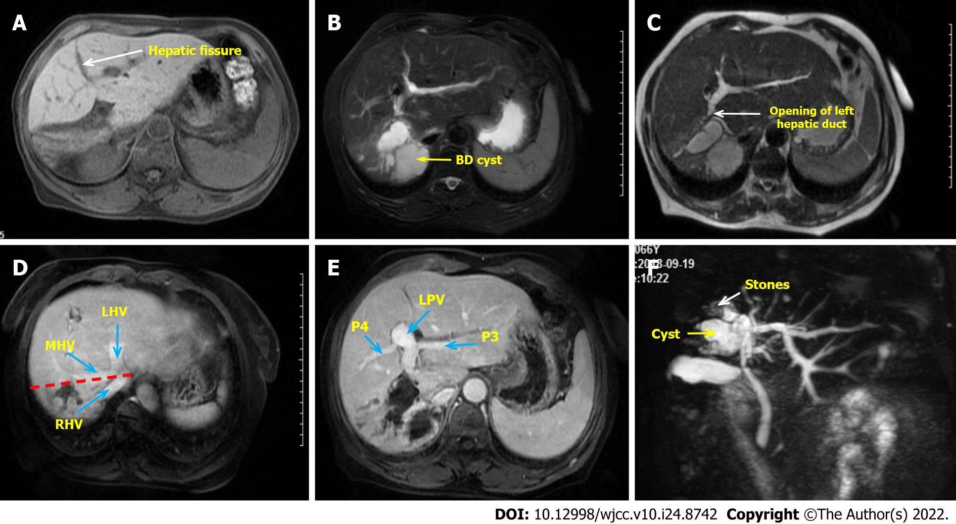
Figure 1 Preoperative clinical image of the abdomen.
A: The right liver atrophy and compensatory hypertrophy of the left liver were seen, with the hepatic fissure (the white arrow) transposed to the right; B: Right hepatic duct cystic dilatation; C: The opening of left hepatic duct (the white arrow) is very close to the cyst; D: The red line shows the expected excision line and the blue arrows show the three major hepatic veins; E: No vascular variation was found and the left portal veins were also transposed to the right; F: Magnetic resonance cholangiopancreatography was used to evaluate intrahepatic and extrahepatic bile ducts. The end of the right hepatic duct is full of stones (the white arrow). LHV: Left hepatic vein; MHV: Middle hepatic vein; P3: Portal vein of Segment 3; P5: Portal vein of Segment 5; RHV: Right hepatic vein.
- Citation: Zhao J, Dang YL. When should endovascular gastrointestinal anastomosis transection Glissonean pedicle not be used in hepatectomy? A case report. World J Clin Cases 2022; 10(24): 8742-8748
- URL: https://www.wjgnet.com/2307-8960/full/v10/i24/8742.htm
- DOI: https://dx.doi.org/10.12998/wjcc.v10.i24.8742


