Copyright
©The Author(s) 2021.
World J Clin Cases. Mar 16, 2021; 9(8): 1996-2000
Published online Mar 16, 2021. doi: 10.12998/wjcc.v9.i8.1996
Published online Mar 16, 2021. doi: 10.12998/wjcc.v9.i8.1996
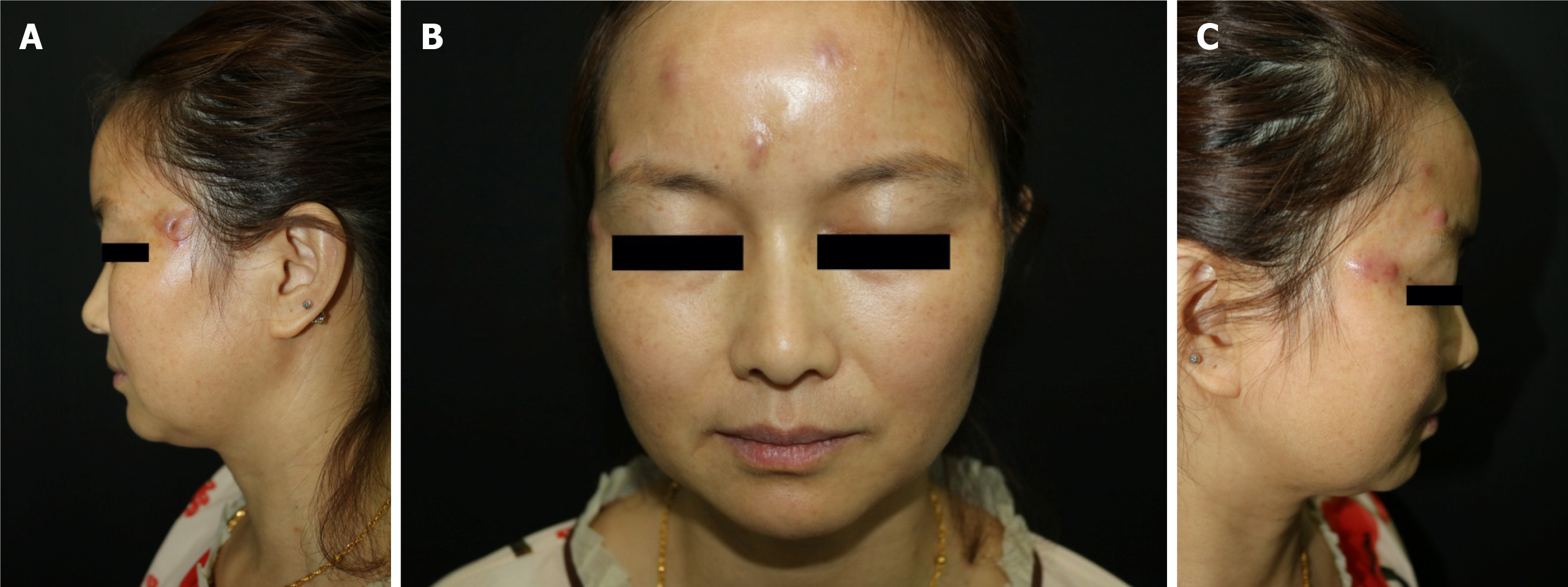
Figure 1 Abscesses and nodules over the argireline injection sites.
A: An abscess with a diameter of approximately 1.0 cm, following an incision, can be seen on the left temple; B: Three red nodules and abscesses with a diameter of approximately 0.5 cm can be seen on the forehead; C: Four red nodules and abscesses with a diameter of approximately 0.5 cm can be seen on the right temple.
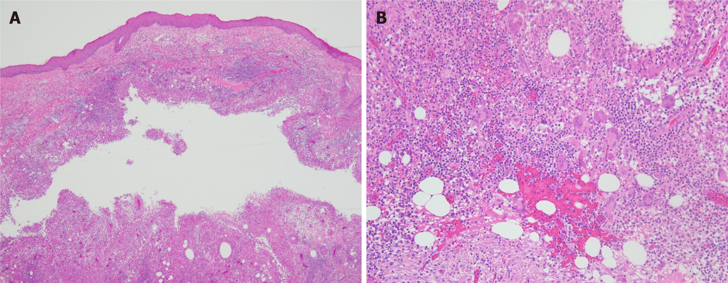
Figure 2 Pathological biopsy of the lesion was performed.
A: A cystic cavity can be seen in the dermis, surrounded by granulomatous structures formed by histiocytes [Hematoxylin-eosin staining (HE) × 40]; B: Scattered around the cystic cavity are multinucleate giant cells, accompanied by lymphocyte, plasma cell, and neutrophil infiltration (HE × 200).
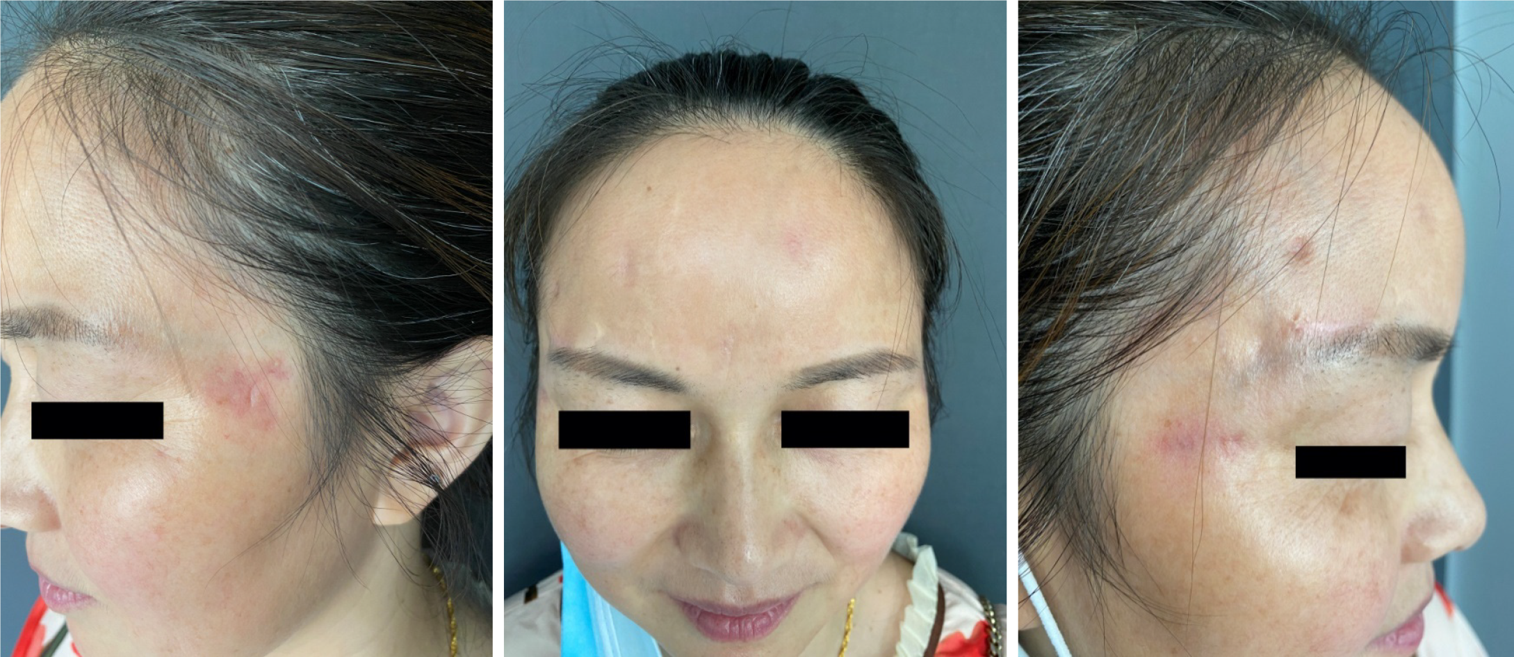
Figure 3 There was clinical resolution after 5 mo of treatment, but scars and pigmentation remained.
- Citation: Chen CF, Liu J, Wang SS, Yao YF, Yu B, Hu XP. Mycobacterium abscessus infection after facial injection of argireline: A case report. World J Clin Cases 2021; 9(8): 1996-2000
- URL: https://www.wjgnet.com/2307-8960/full/v9/i8/1996.htm
- DOI: https://dx.doi.org/10.12998/wjcc.v9.i8.1996


