Copyright
©The Author(s) 2021.
World J Clin Cases. Mar 16, 2021; 9(8): 1976-1982
Published online Mar 16, 2021. doi: 10.12998/wjcc.v9.i8.1976
Published online Mar 16, 2021. doi: 10.12998/wjcc.v9.i8.1976
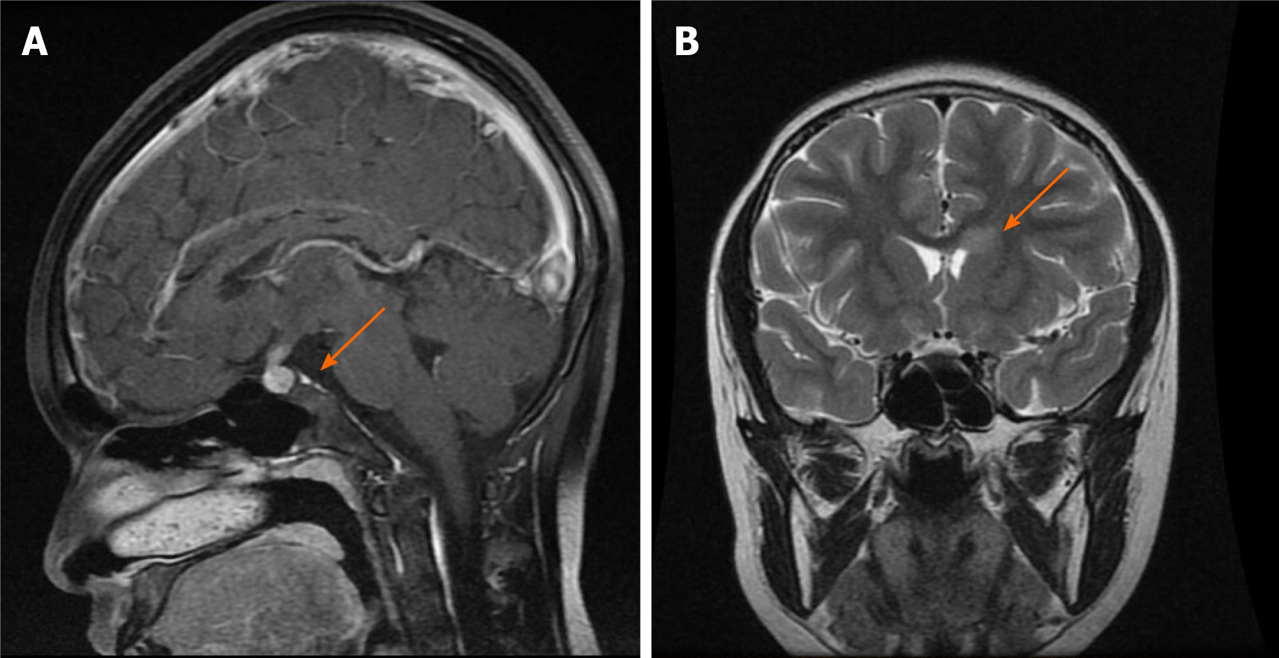
Figure 1 Magnetic resonance imaging of the brain.
A: A sellar mass with thickness of the pituitary stalk and absence of the hyperintensity signal of the posterior pituitary; B: Abnormal enhancement shadow in the left anterior horn of the ventricle.
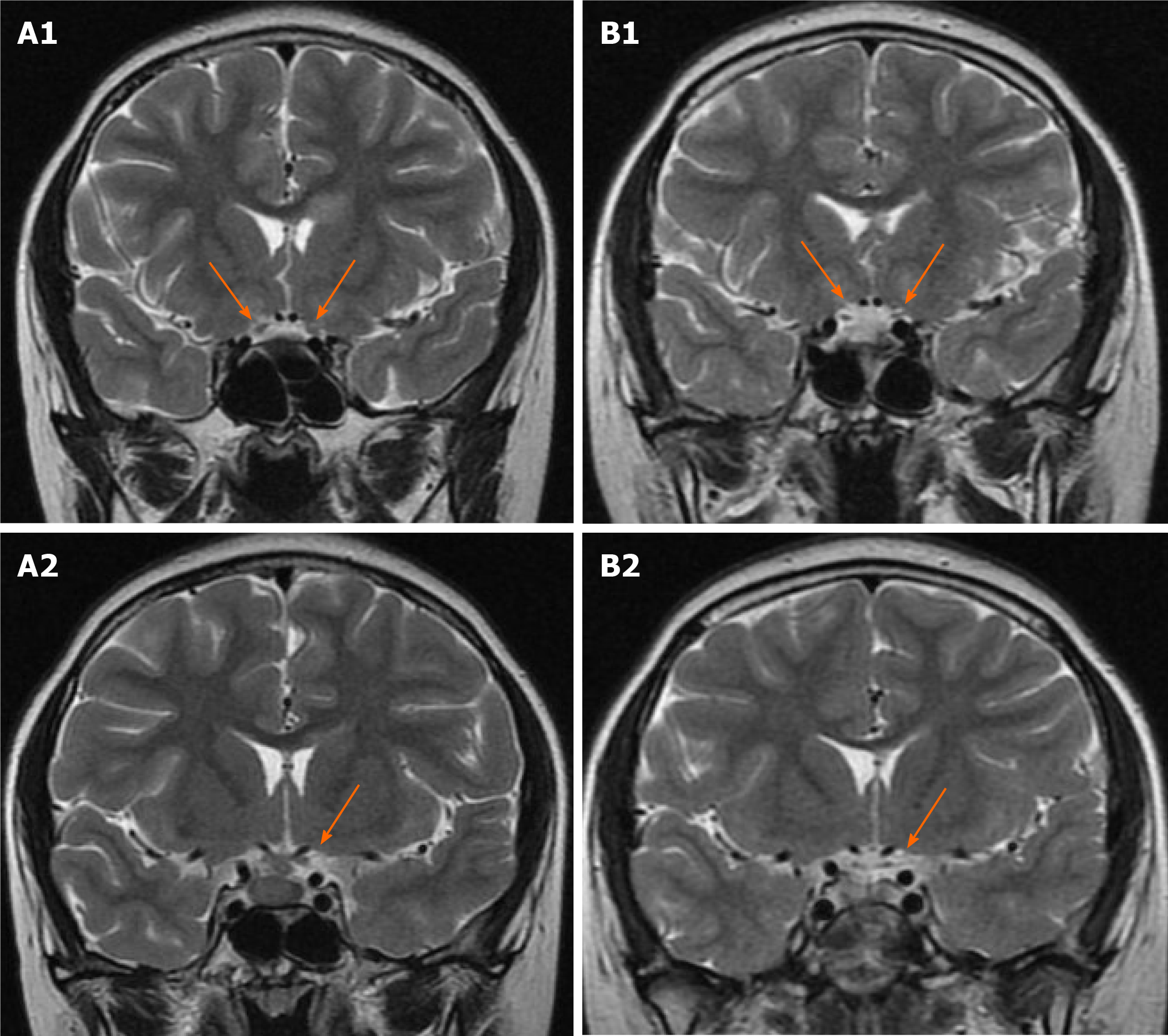
Figure 2 Magnetic resonance imaging of the optic nerve.
A: Pretreatment image showing enlarged optic nerve; B: Image after radiotherapy and chemotherapy showing atrophy of the optic nerve.
- Citation: Yang N, Zhu HJ, Yao Y, He LY, Li YX, You H, Zhang HB. Diabetes insipidus with impaired vision caused by germinoma and perioptic meningeal seeding: A case report. World J Clin Cases 2021; 9(8): 1976-1982
- URL: https://www.wjgnet.com/2307-8960/full/v9/i8/1976.htm
- DOI: https://dx.doi.org/10.12998/wjcc.v9.i8.1976


