Copyright
©The Author(s) 2021.
World J Clin Cases. Mar 16, 2021; 9(8): 1901-1908
Published online Mar 16, 2021. doi: 10.12998/wjcc.v9.i8.1901
Published online Mar 16, 2021. doi: 10.12998/wjcc.v9.i8.1901
Figure 1 Bronchoscopy findings.
A-C: Bronchoscopy images of main trachea (A), left main bronchus (B) and opening of the right basal segmental bronchus (C) showed that the bronchi were patent, and sporadic mucosal petechiae and bloody sputum were seen in the bilateral bronchi.
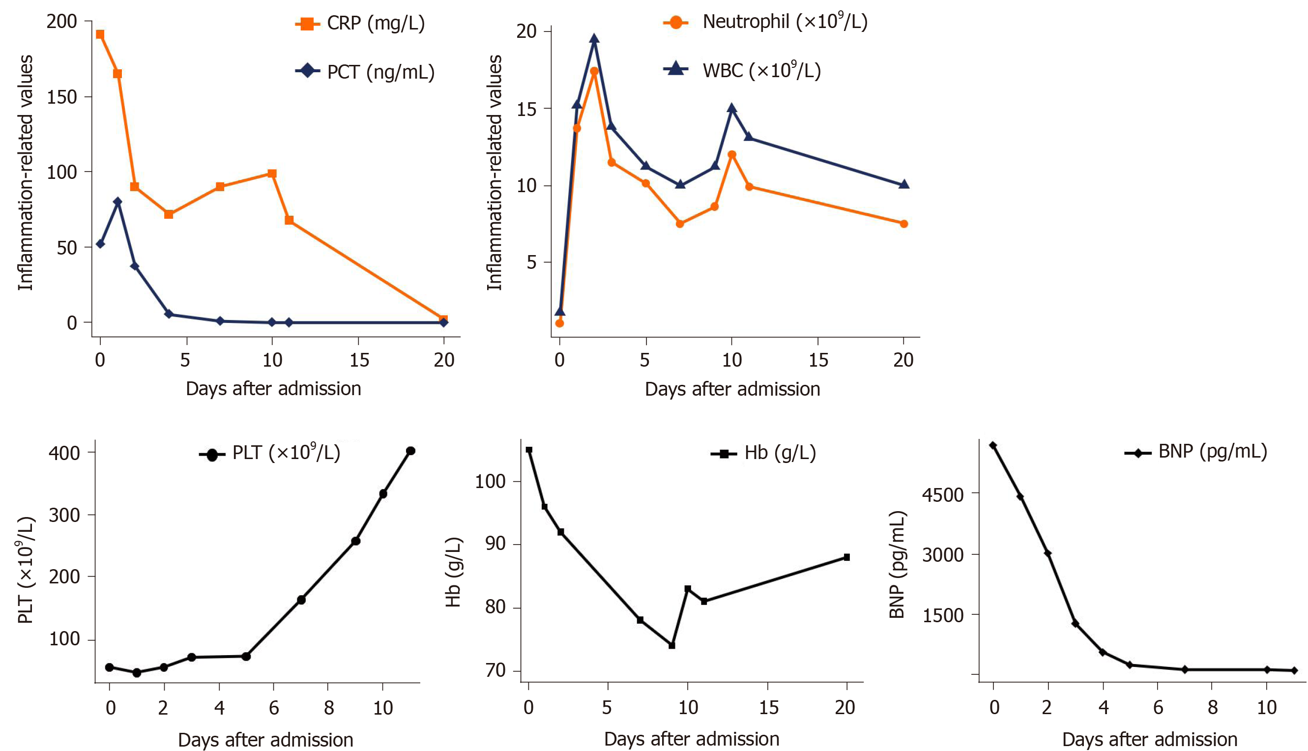
Figure 2 Changes in laboratory parameters after admission.
In the first 5 d of antiviral treatment after admission: inflammatory values were elevated; platelets and hemoglobin were decreased consistent with hemorrhagic complications; and elevated brain natriuretic peptide indicated the existence of myocardial impairment. After 5 d, penicillin was administered and the above vital parameters improved. WBC: White blood cell; CRP: C-reactive protein; PCT: Procalcitonin; PLT: Platelets; Hb: Hemoglobin; BNP: Brain natriuretic peptide.
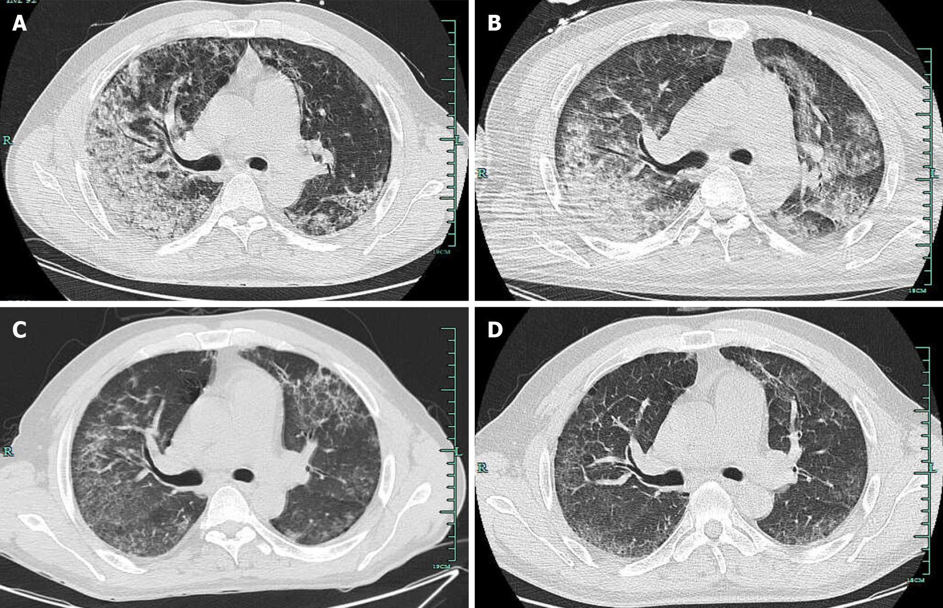
Figure 3 Computed tomography findings.
A-D: Computed tomography images were respectively taken on admission (A), 1 wk after admission (B), 1 wk before discharge (C), and 3 wk after discharge (D). Gradual resolution of inflammation in the bilateral lungs was seen during the treatment course.
- Citation: Bao QH, Yu L, Ding JJ, Chen YJ, Wang JW, Pang JM, Jin Q. Severe community-acquired pneumonia caused by Leptospira interrogans: A case report and review of literature. World J Clin Cases 2021; 9(8): 1901-1908
- URL: https://www.wjgnet.com/2307-8960/full/v9/i8/1901.htm
- DOI: https://dx.doi.org/10.12998/wjcc.v9.i8.1901


