Copyright
©The Author(s) 2021.
World J Clin Cases. Feb 6, 2021; 9(4): 801-811
Published online Feb 6, 2021. doi: 10.12998/wjcc.v9.i4.801
Published online Feb 6, 2021. doi: 10.12998/wjcc.v9.i4.801
Figure 1 Guidance by ultrasound and digital subtraction angiography.
A: The dilated bile duct was punctured with an 18G cannula puncture needle; B: A guide wire was introduced through the puncture needle sheath; C: Two guide wires were introduced through the 8F catheter; D: One guide wire was introduced into the stent conveyor, and the other into the 8F catheter, ready to push the iodine-125 (125I) seed strands; E: Successful placement of stent and 125I seed strands; F: External drainage tube angiography showing the unobstructed stent; G: 125I seeds were placed into 4F plastic tubes to make 125I seed strands.
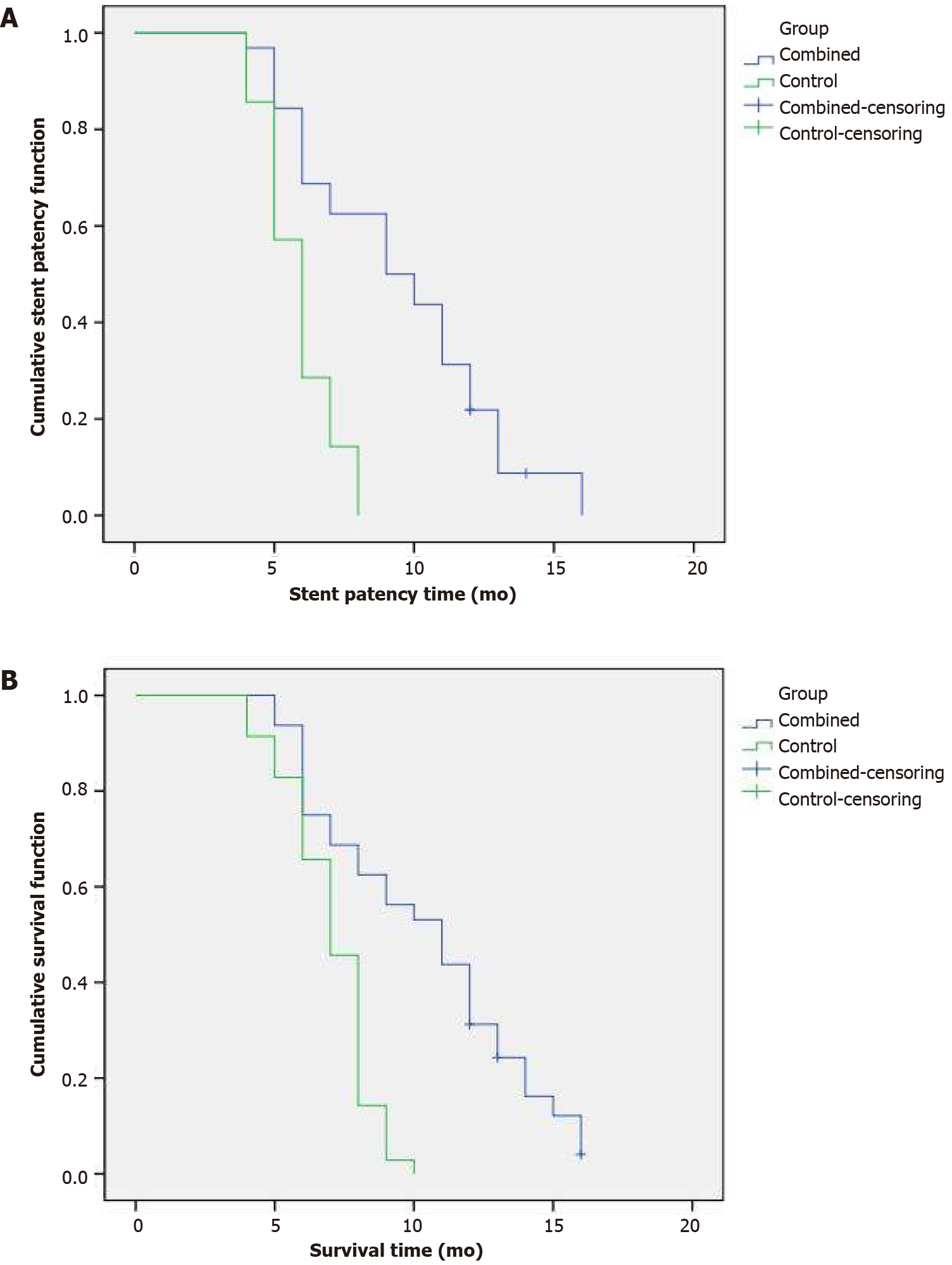
Figure 2 Median stent patency times and survival time for the combined group.
A: The survival curve showed that the median stent patency time in the combined group was 9.000 ± 1.414 mo (95% confidence interval (CI): 6.228-11.772 mo), the control group was 6.000 ± 0.267 mo (95%CI: 5.476-6.524 mo), and there was a significant difference (P = 0.000); B: The survival curve showed that the median survival time of the combined group was 11.000 ± 1.403 mo (95%CI: 8.250-13.750 mo), the control group was 7.000 ± 0.327 mo (95%CI: 6.358-7.642 mo), and there was a significant difference between the two groups (P = 0.000).
- Citation: Wang HW, Li XJ, Li SJ, Lu JR, He DF. Biliary stent combined with iodine-125 seed strand implantation in malignant obstructive jaundice. World J Clin Cases 2021; 9(4): 801-811
- URL: https://www.wjgnet.com/2307-8960/full/v9/i4/801.htm
- DOI: https://dx.doi.org/10.12998/wjcc.v9.i4.801


