Copyright
©The Author(s) 2021.
World J Clin Cases. Nov 26, 2021; 9(33): 10374-10381
Published online Nov 26, 2021. doi: 10.12998/wjcc.v9.i33.10374
Published online Nov 26, 2021. doi: 10.12998/wjcc.v9.i33.10374
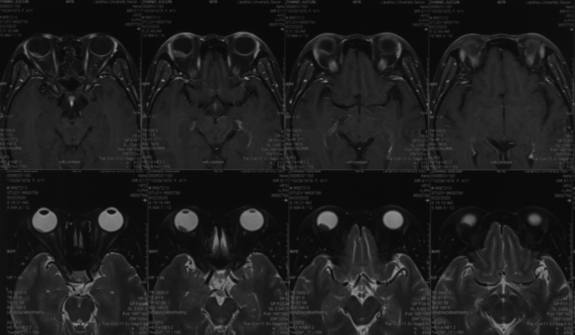
Figure 1 Magnetic resonance imaging of the right eye showed an abnormal signal shadow behind the right eye bulb, short T1 and T2 signals, and high diffusion-weighted imaging signals.
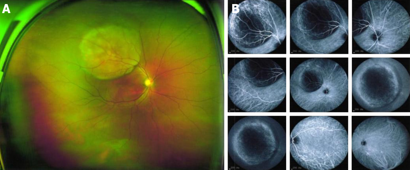
Figure 2 Ophthalmoscopy of the right eye.
A, B: Solid dark gray mass in the posterior segment (choroid) with intense brown pigmentation, occupying posterior third of the vitreous chamber along with mild retinal detachment observed at the peripheral rim of the nodular choroidal mass. The mass size of 11.11 mm × 12.1 mm.
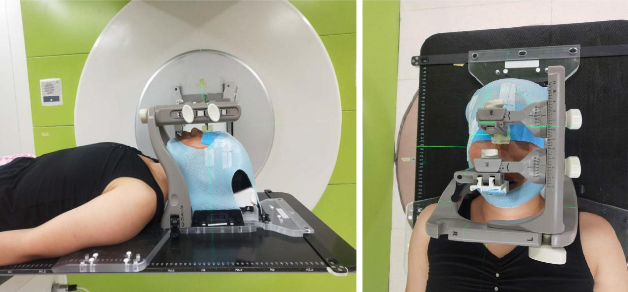
Figure 3 Head immobilization with trUpoint.
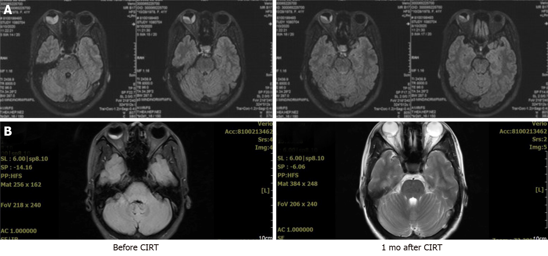
Figure 4 Images after 5 fractions of carbon ion radiotherapy, and 1 mo after carbon ion radiotherapy.
A: The tumor is bigger, retinal detachment was obvious; B: The choroidal tumor of right eye was smaller and retinal detachment was improved. CIRT: Carbon ion radiotherapy.
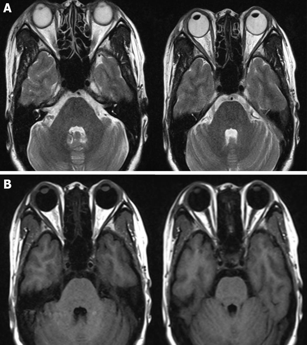
Figure 5 Choroidal tumor of the right eye disappeared completely 3 mo after carbon ion radiotherapy.
A: T2W magnetic resonance imaging (MRI); B: T1W MRI.
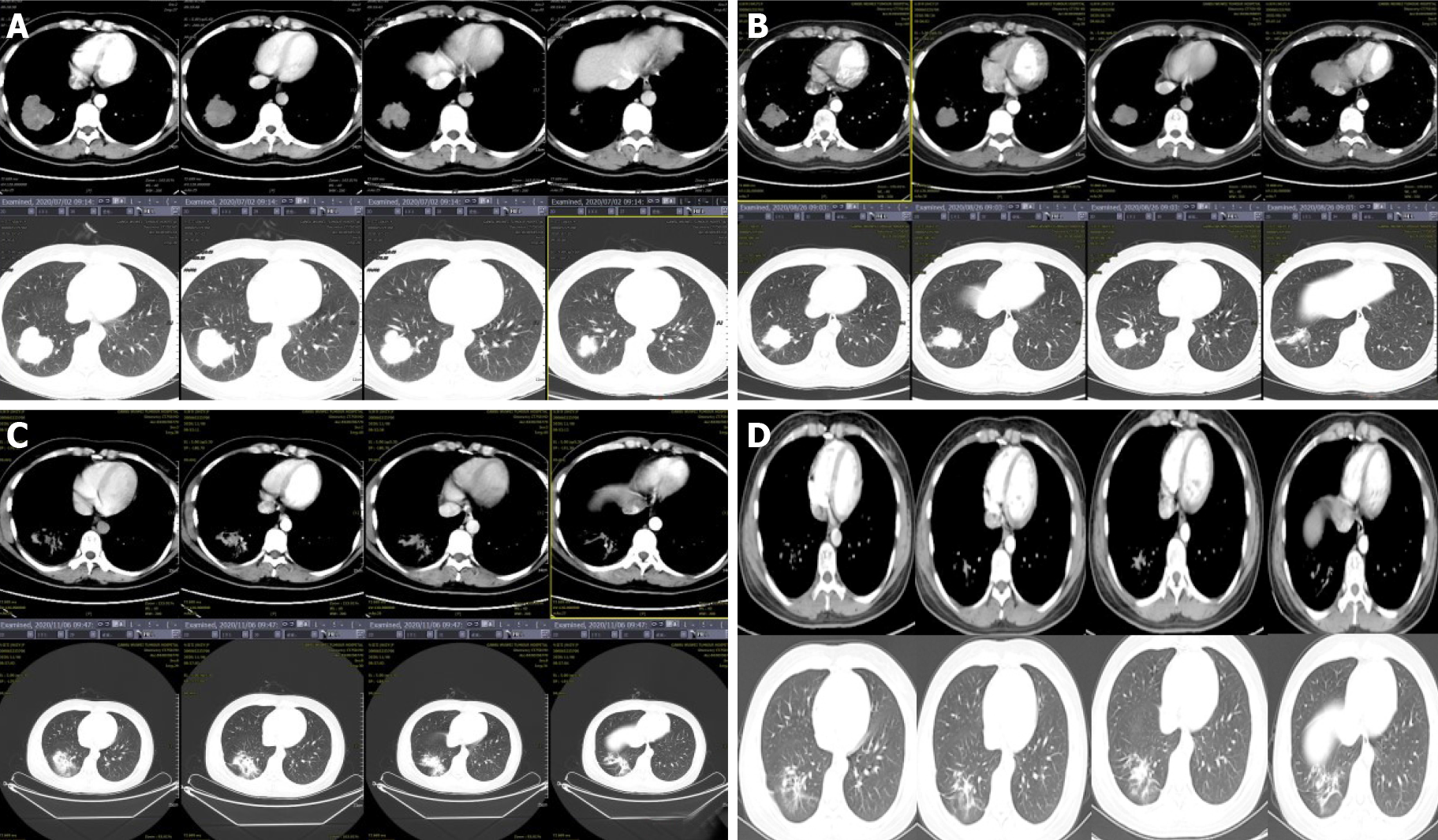
Figure 6 Images of lung tumor.
A: Size of the lung tumor before carbon ion radiotherapy (CIRT) was 4.7 cm × 5.2 cm; B: Size of the lung tumor 1 mo after CIRT was 4.2 cm × 3.5 cm; C: At 3 mo after CIRT, the right lung tumor size was 3.6 cm × 2.5 cm; D: At 7 mo, the lung tumor disappeared completely, only showing a little pulmonary fibrosis.
- Citation: Zhang YS, Hu TC, Ye YC, Han JH, Li XJ, Zhang YH, Chen WZ, Chai HY, Pan X, Wang X, Yang YL. Carbon ion radiotherapy for synchronous choroidal melanoma and lung cancer: A case report. World J Clin Cases 2021; 9(33): 10374-10381
- URL: https://www.wjgnet.com/2307-8960/full/v9/i33/10374.htm
- DOI: https://dx.doi.org/10.12998/wjcc.v9.i33.10374


