Copyright
©The Author(s) 2021.
World J Clin Cases. Nov 26, 2021; 9(33): 10126-10133
Published online Nov 26, 2021. doi: 10.12998/wjcc.v9.i33.10126
Published online Nov 26, 2021. doi: 10.12998/wjcc.v9.i33.10126
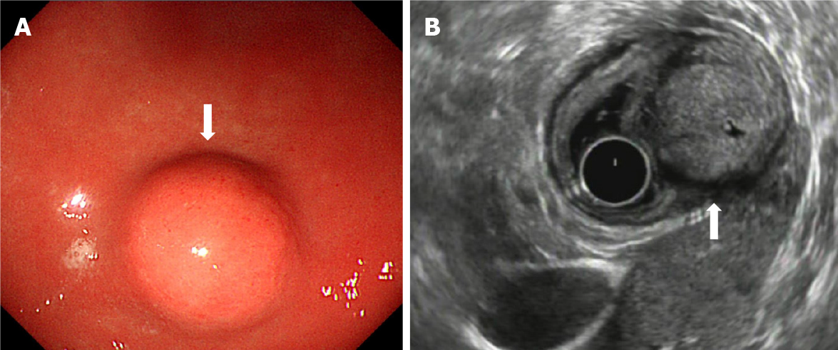
Figure 1 Endoscopic view of a well-circumscribed elevated lesion with normal overlying mucosa at the stomach antrum (arrow).
A: Endoscopic ultrasound (EUS) characteristics of glomus tumors; B: Tumor originates from the fourth EUS layer (muscularis propria) and shows slight hypoechogenicity and a characteristic marginal halo around the tumor.
Figure 2 Computed tomography features of glomus tumors.
Contrast-enhanced computed tomography image obtained during the plain scan, arterial phase, portal phase; homogeneous strong enhancement is seen in the gastric antrum lesion (arrow).
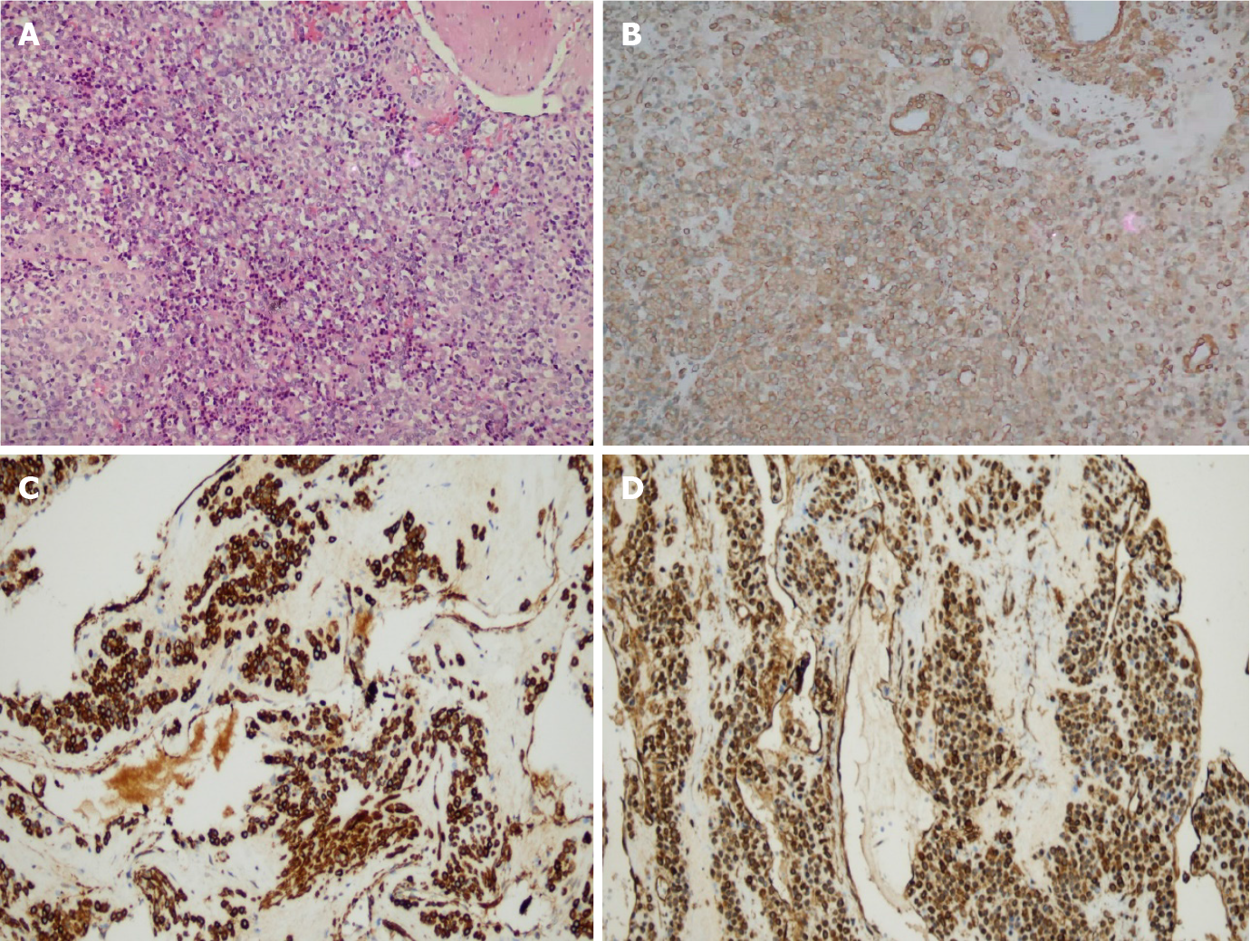
Figure 3 Photomicrograph of the tumor cells.
A: Clusters of uniform, round cells around dilated blood vessels, cytoplasm was transparent or reddish, and the cells had no atypia or mitotic appearance; B: Positive smooth muscle actin staining of tumor cells, which shows a brown cytoplasmic stain; C: Positive h-caldesmon staining of tumor cells, which shows a brown cytoplasmic stain; D: Positive vimentin staining of tumor cells, which shows a brown cytoplasmic stain. Hematoxylin-eosin; original magnification: 200 ×.
- Citation: Bai B, Mao CS, Li Z, Kuang SL. Endoscopic ultrasonography diagnosis of gastric glomus tumors. World J Clin Cases 2021; 9(33): 10126-10133
- URL: https://www.wjgnet.com/2307-8960/full/v9/i33/10126.htm
- DOI: https://dx.doi.org/10.12998/wjcc.v9.i33.10126


