Copyright
©The Author(s) 2021.
World J Clin Cases. Nov 16, 2021; 9(32): 10024-10032
Published online Nov 16, 2021. doi: 10.12998/wjcc.v9.i32.10024
Published online Nov 16, 2021. doi: 10.12998/wjcc.v9.i32.10024
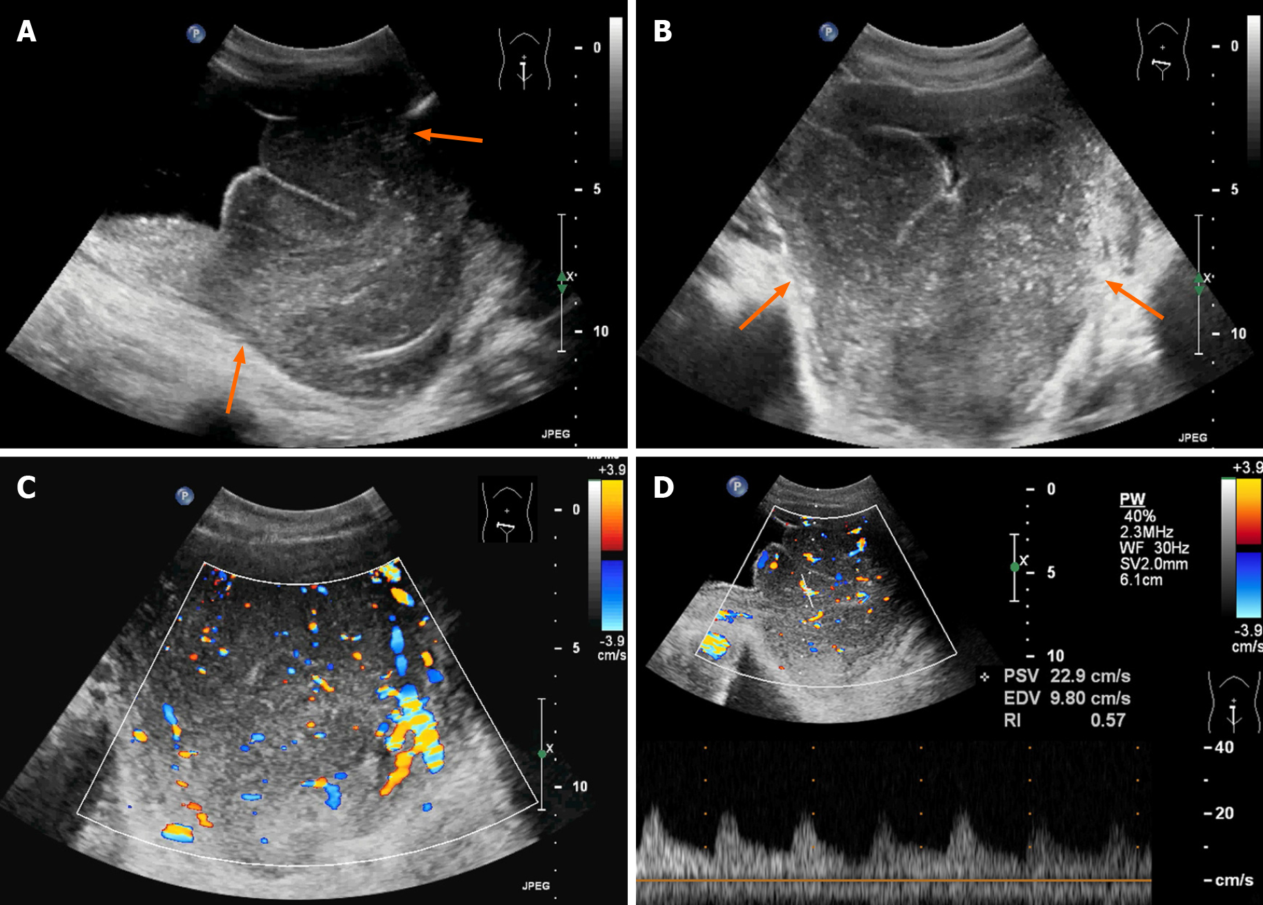
Figure 1 Ultrasonographic findings of the lesion.
A and B: A large homogeneous hypoechoic mass was detected in the bladder (arrows), with an unclear boundary and irregular shape. There were fine linear echogenic strands distributed inside; C: Color Doppler flow imaging showed that there were rich blood flow signals in the mass; D: Spectral Doppler ultrasound imaging showed the artery spectrum detected within the mass (Vmax = 22.9 cm/s, resistance index: 0.57).
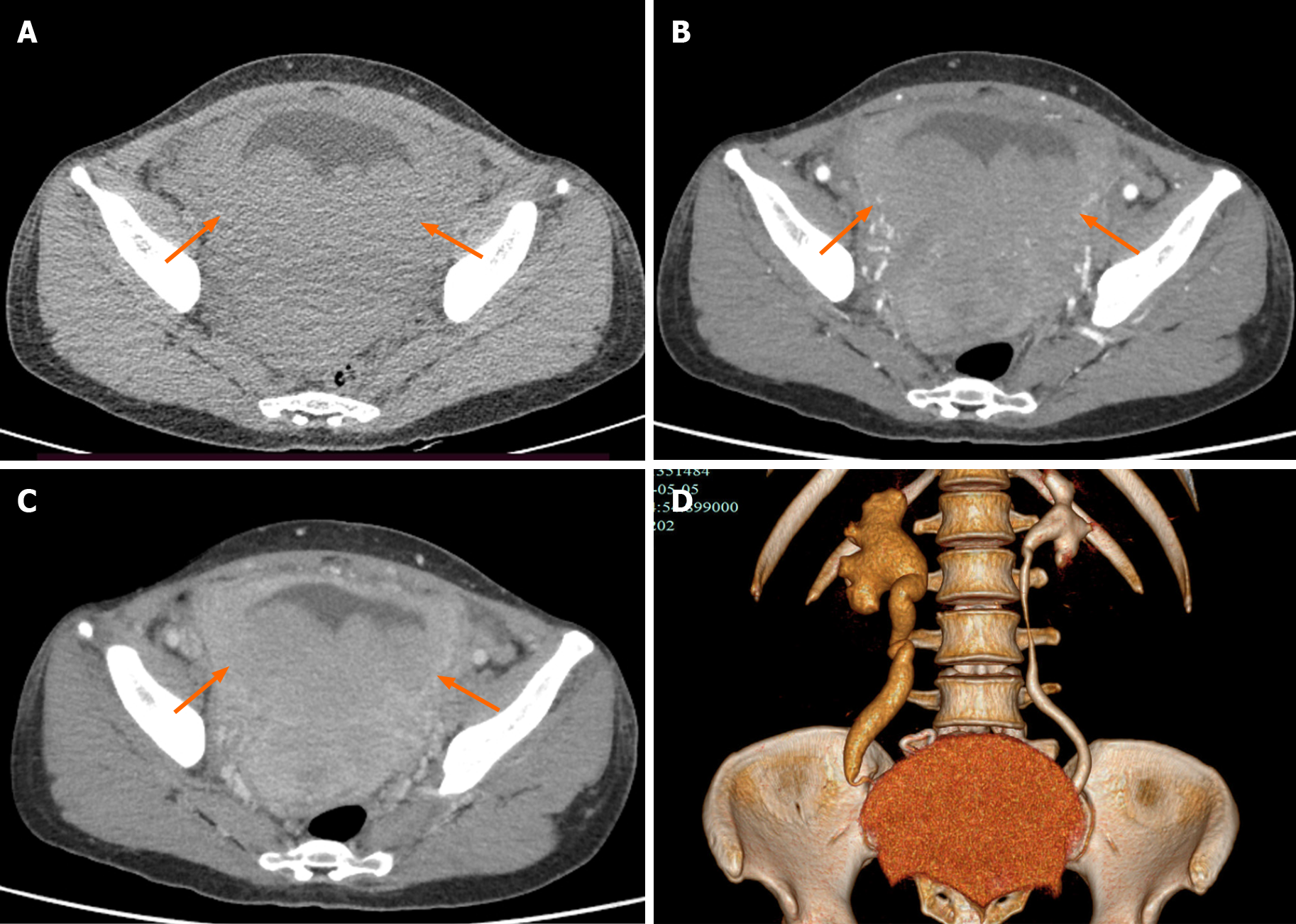
Figure 2 Computed tomography findings of the lesion.
A: The bladder wall was thickened, and there were multiple low-density nodules in the bladder with unclear boundaries and irregular morphologies (arrow); B and C: Continuous enhancement of the mass was observed; D: The ureter and renal pelvis area of the right kidney was dilated.
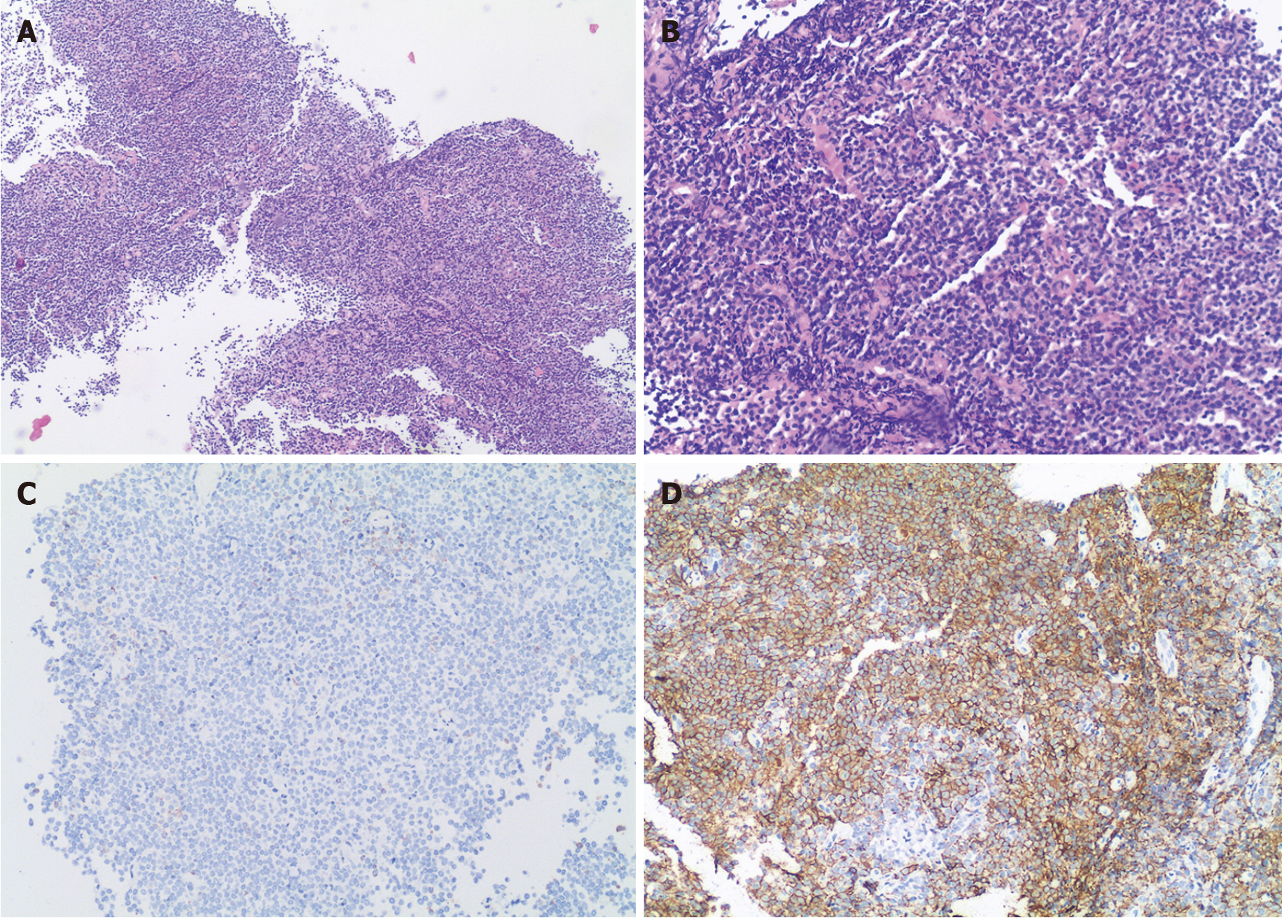
Figure 3 Histopathological findings of the mass tissues.
There was diffuse infiltration of atypical lymphoid cells. A: Hematoxylin and eosin (HE) staining, magnification 40 ×; B: HE staining, magnification 100 ×; C: Immunohistochemical (IHC) staining, negative for CD3, magnification 100 ×; D: IHC staining, positive for CD20, magnification 100 ×.
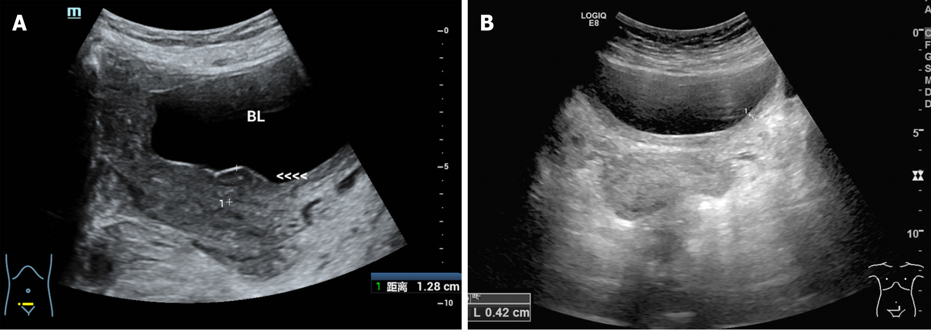
Figure 4 Ultrasonographic findings during the chemotherapy.
A: After two cycles of chemotherapy, the mass had disappeared with localized thickening (13 mm) of the bladder wall; B: After three cycles of chemotherapy, bladder wall thickness was only 4 mm, which indicated excellent therapeutic response to the treatment.
- Citation: Jiang ZZ, Zheng YY, Hou CL, Liu XT. Primary mucosal-associated lymphoid tissue extranodal marginal zone lymphoma of the bladder from an imaging perspective: A case report. World J Clin Cases 2021; 9(32): 10024-10032
- URL: https://www.wjgnet.com/2307-8960/full/v9/i32/10024.htm
- DOI: https://dx.doi.org/10.12998/wjcc.v9.i32.10024


