Copyright
©The Author(s) 2021.
World J Clin Cases. Nov 6, 2021; 9(31): 9607-9616
Published online Nov 6, 2021. doi: 10.12998/wjcc.v9.i31.9607
Published online Nov 6, 2021. doi: 10.12998/wjcc.v9.i31.9607
Figure 1 Typical lymphoma cells were found in bone marrow cytology.
Magnification of images was 100 × 10.
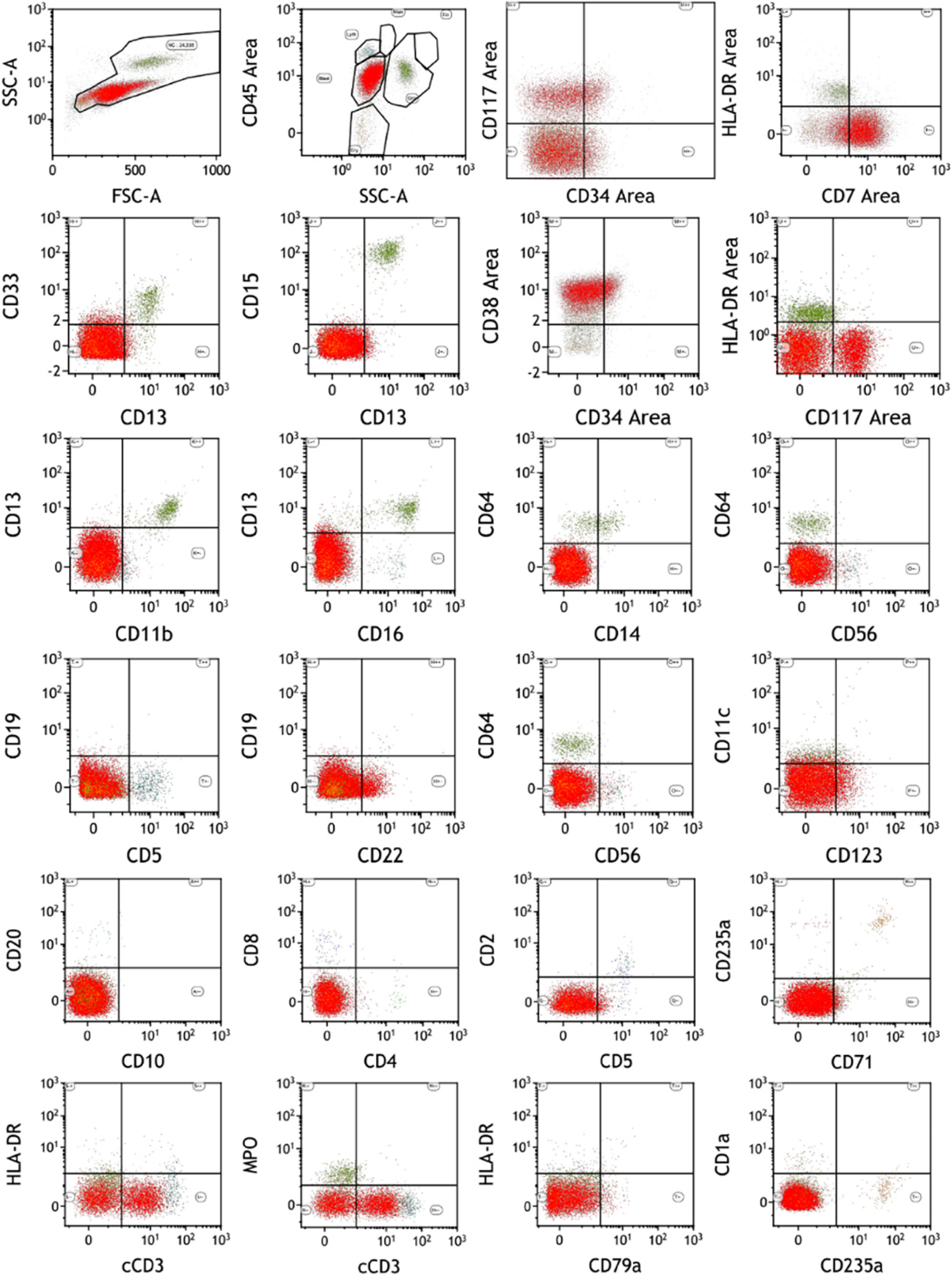
Figure 2 Early T-cell precursor acute lymphoblastic leukemia was definitely diagnosed by flow immunotyping.
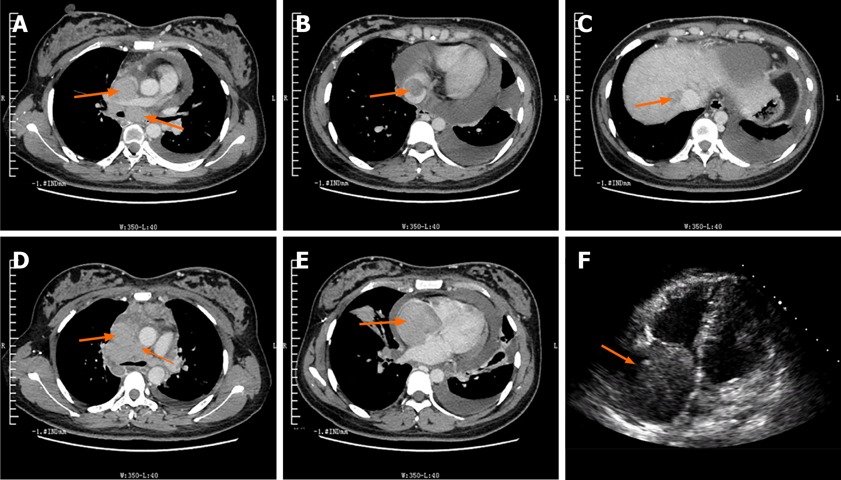
Figure 3 Chest enhanced computed tomography and ultrasonic cardiogram showed extensive thrombi and heart thrombosis.
A: Low-density filling defect in the superior vena cava; arrow indicate enlarged lymph nodes under the carina of the trachea; B: Low-density filling defect (arrow) at the proximal end of the inferior vena cava; C: Low-density filling defect (arrow) in right hepatic veins; D: Multiple swollen and fused lymph nodes in the anterior trachea; arrow indicates the low-density filling defect in the superior vena cava; E: Low-density filling defect (arrow) in the right atrium; note the lack of infiltration of adjacent structures; F: Cardiac thrombus (arrow) in the right atrium in echocardiography imaging.
- Citation: Ma YY, Zhang QC, Tan X, Zhang X, Zhang C. T-cell lymphoblastic lymphoma with extensive thrombi and cardiac thrombosis: A case report and review of literature. World J Clin Cases 2021; 9(31): 9607-9616
- URL: https://www.wjgnet.com/2307-8960/full/v9/i31/9607.htm
- DOI: https://dx.doi.org/10.12998/wjcc.v9.i31.9607


