Copyright
©The Author(s) 2021.
World J Clin Cases. Oct 26, 2021; 9(30): 9236-9243
Published online Oct 26, 2021. doi: 10.12998/wjcc.v9.i30.9236
Published online Oct 26, 2021. doi: 10.12998/wjcc.v9.i30.9236
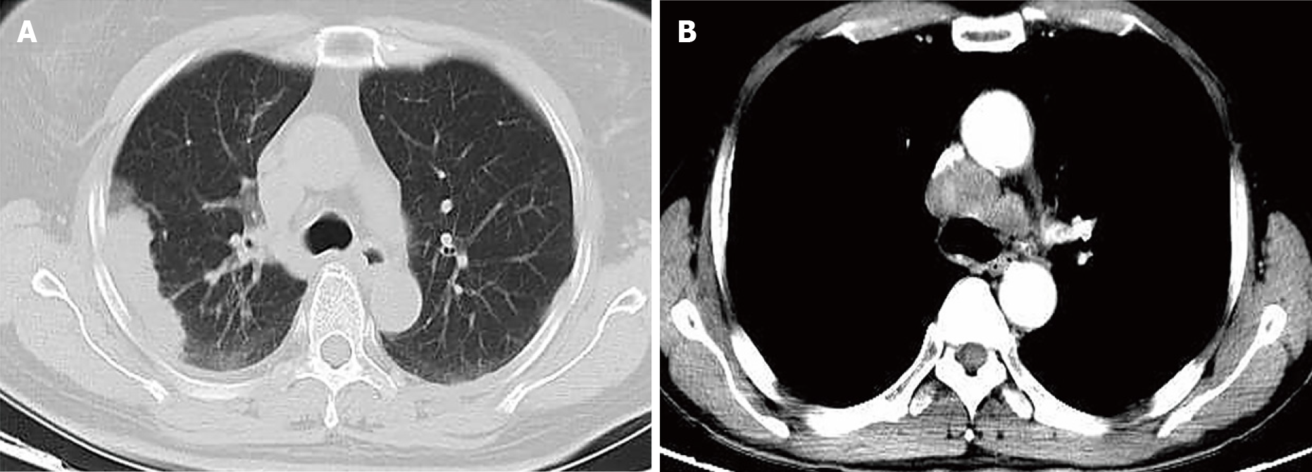
Figure 1 Chest computed tomography results of patients with primary pulmonary enteric adenocarcinoma.
A: Case 4 with pulmonary enteric adenocarcinoma (PEAC) whose lesion was located in the right upper lobe; B: Case 1 with PEAC whose large lesion was located in the right lung, with mediastinal lymph node metastasis.
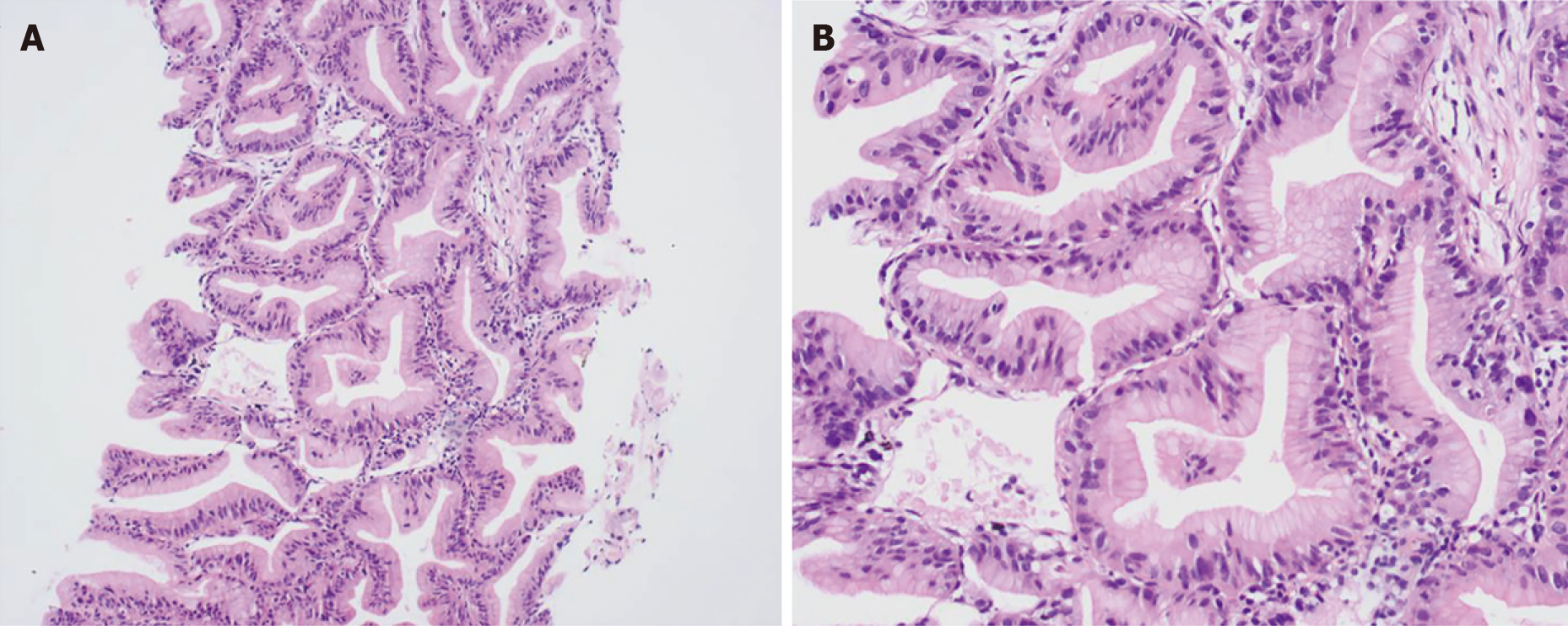
Figure 2 Pathology of case 6 with pulmonary enteric adenocarcinoma (HE staining).
A: × 100; B: × 400.
- Citation: Tu LF, Sheng LY, Zhou JY, Wang XF, Wang YH, Shen Q, Shen YH. Diagnosis and treatment of primary pulmonary enteric adenocarcinoma: Report of Six cases. World J Clin Cases 2021; 9(30): 9236-9243
- URL: https://www.wjgnet.com/2307-8960/full/v9/i30/9236.htm
- DOI: https://dx.doi.org/10.12998/wjcc.v9.i30.9236


