Copyright
©The Author(s) 2021.
World J Clin Cases. Oct 26, 2021; 9(30): 9101-9107
Published online Oct 26, 2021. doi: 10.12998/wjcc.v9.i30.9101
Published online Oct 26, 2021. doi: 10.12998/wjcc.v9.i30.9101
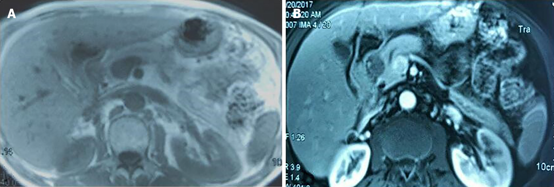
Figure 1 Magnetic resonance imaging findings of pancreatic granular cell tumor.
A: The tumor showed a slight hypointensy on the T1 weighted image; B: The surrounding and center of the tumor were equal signal and hypointense on T2 weighted image, respectively.
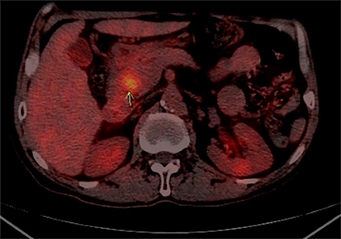
Figure 2
The L1/2 level of the descending part of the duodenum and head of pancreas and soft tissue nodules, and the two is unclear, the computed tomography value is about 45 U, the sectional area of about 24 mm × 22 mm, uptake in the SUV, the maximum value of about 4.
8, two hour delay imaging, radiation higher than before, the maximum value of 5.2 SUV, a visible display of pancreatic duct.
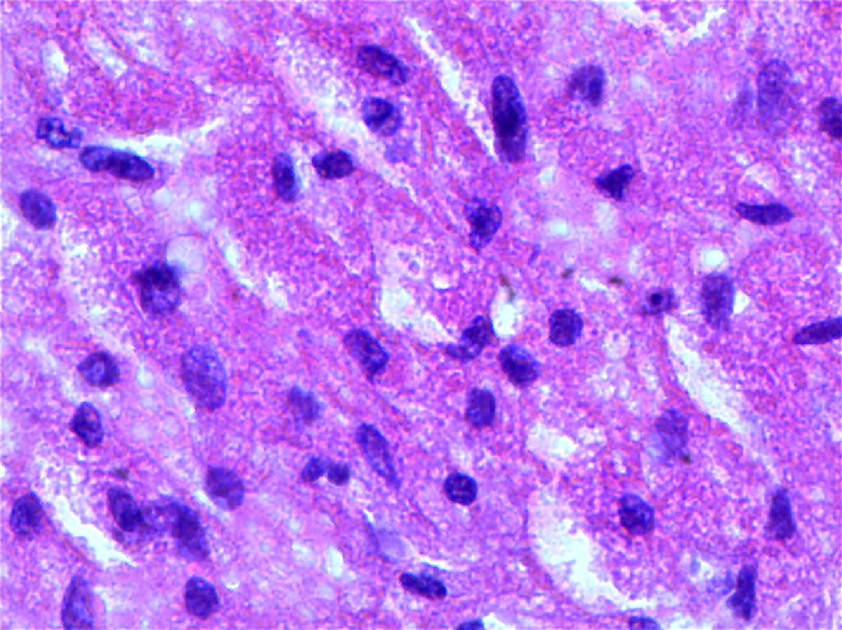
Figure 3
The tumor cells that consisted of a large number cytoplasmiceosinophilic were oval.
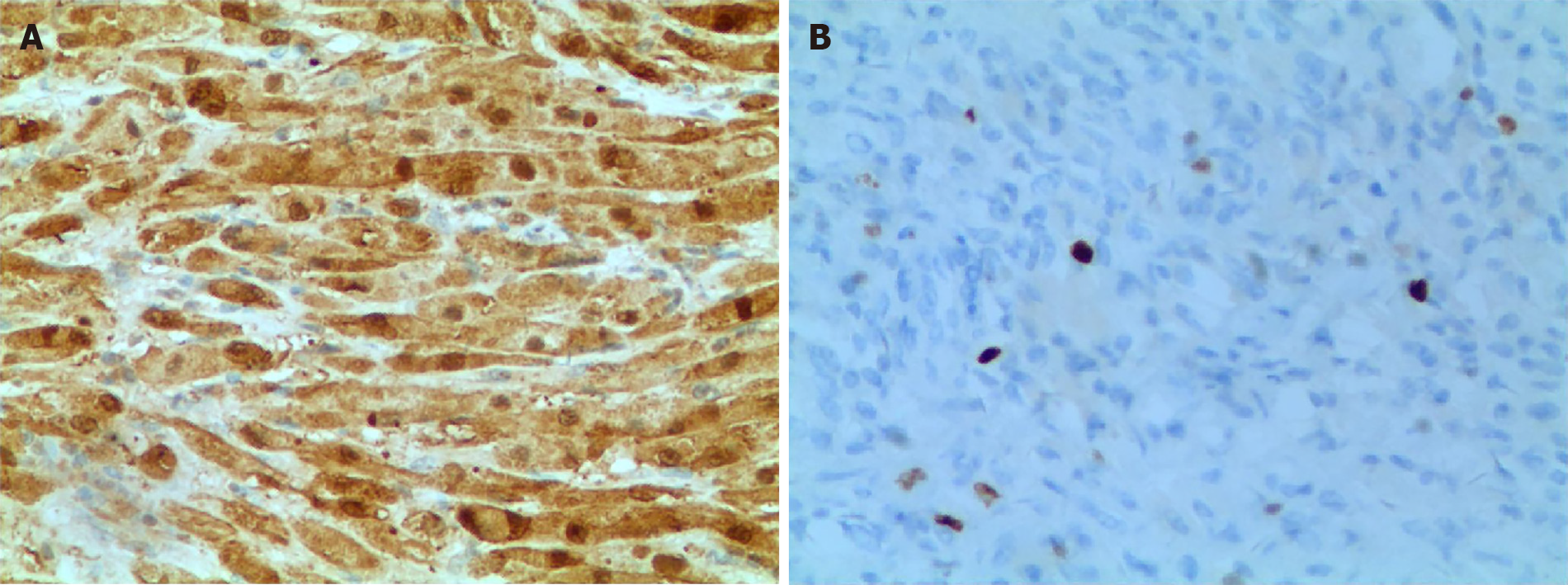
Figure 4 Periodic Acid-Schiff staining and immunohistochemistry showed positive staining of S-100 protein in tumor cells.
A: Periodic Acid-Schiff staining; B: Immunohistochemistry.
- Citation: Zhu MH, Nie CF. Particular tumor of the pancreas: A case report . World J Clin Cases 2021; 9(30): 9101-9107
- URL: https://www.wjgnet.com/2307-8960/full/v9/i30/9101.htm
- DOI: https://dx.doi.org/10.12998/wjcc.v9.i30.9101


