Copyright
©The Author(s) 2021.
World J Clin Cases. Oct 26, 2021; 9(30): 9023-9037
Published online Oct 26, 2021. doi: 10.12998/wjcc.v9.i30.9023
Published online Oct 26, 2021. doi: 10.12998/wjcc.v9.i30.9023
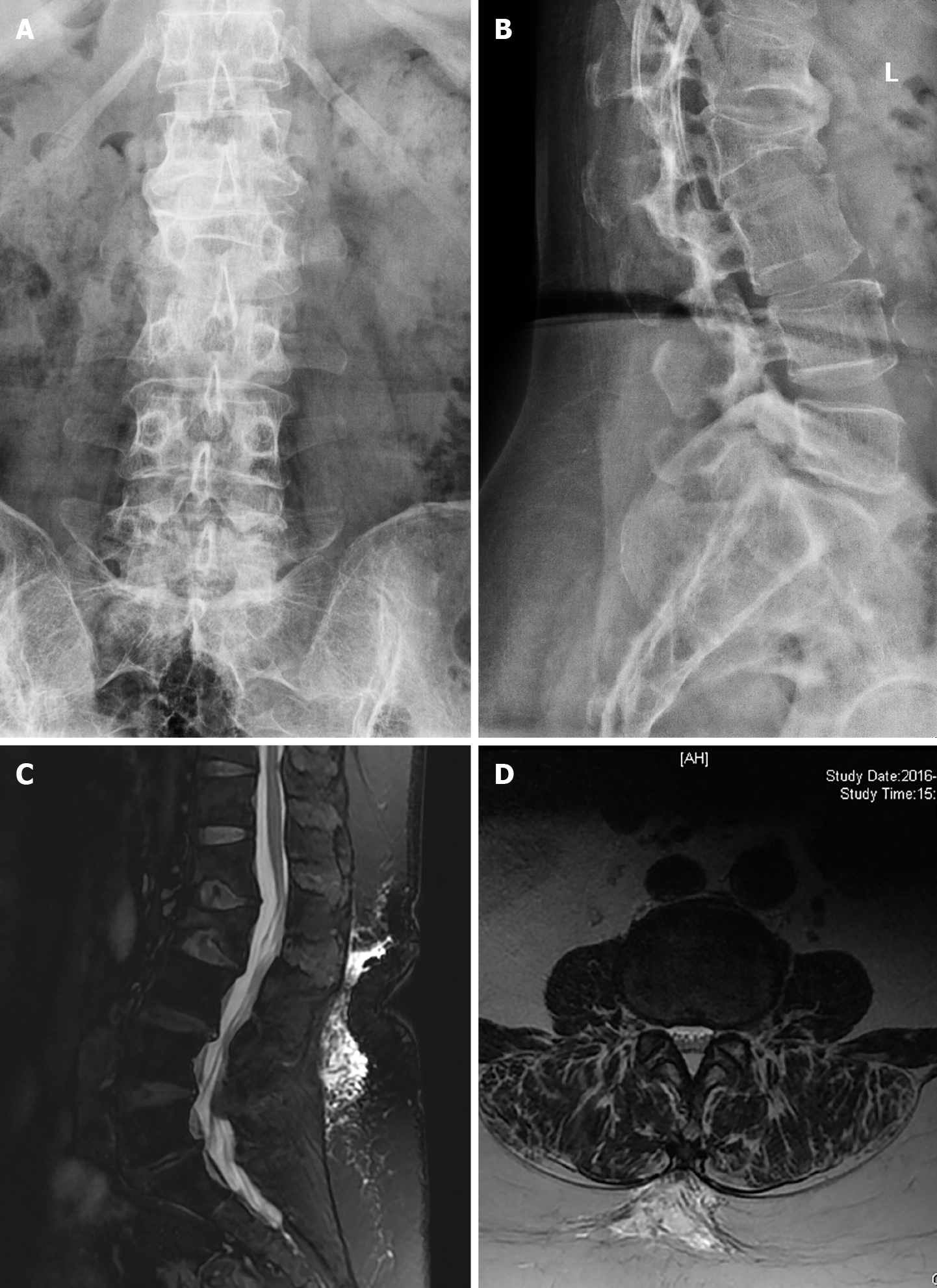
Figure 1 The score indicated “spinal stability”.
A, B: Lumbar spine X-ray image, showing the presence of a physiological curvature; C: A sagittal plane magnetic resonance (MR) in which T11 and L1-2 lesions are visible; D: A normal MR image.
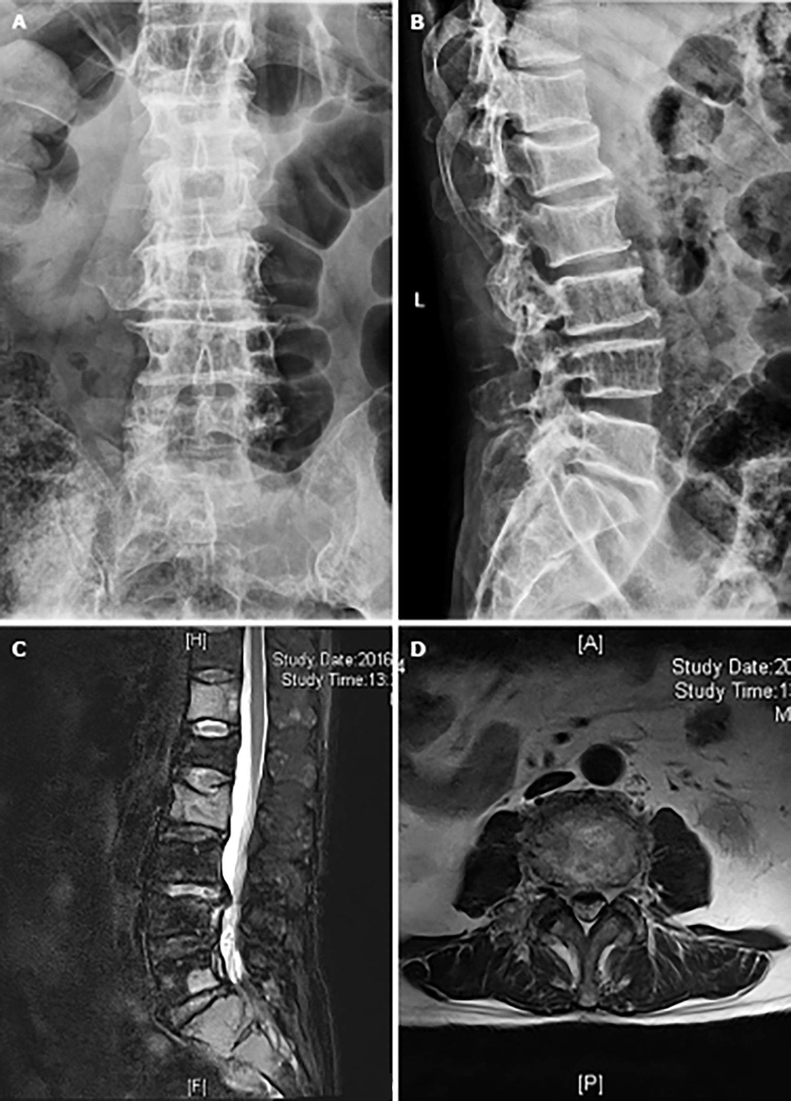
Figure 2 The score indicated “potential instability”.
A, B: Lumbar spine X-ray image showing the presence of a lumbar physiological curvature; C: Sagittal plane magnetic resonance (MR) images in which T12, L2 and S1-2 lesions are visible; D: Lumbar disc herniation on a cross-sectional MR image.
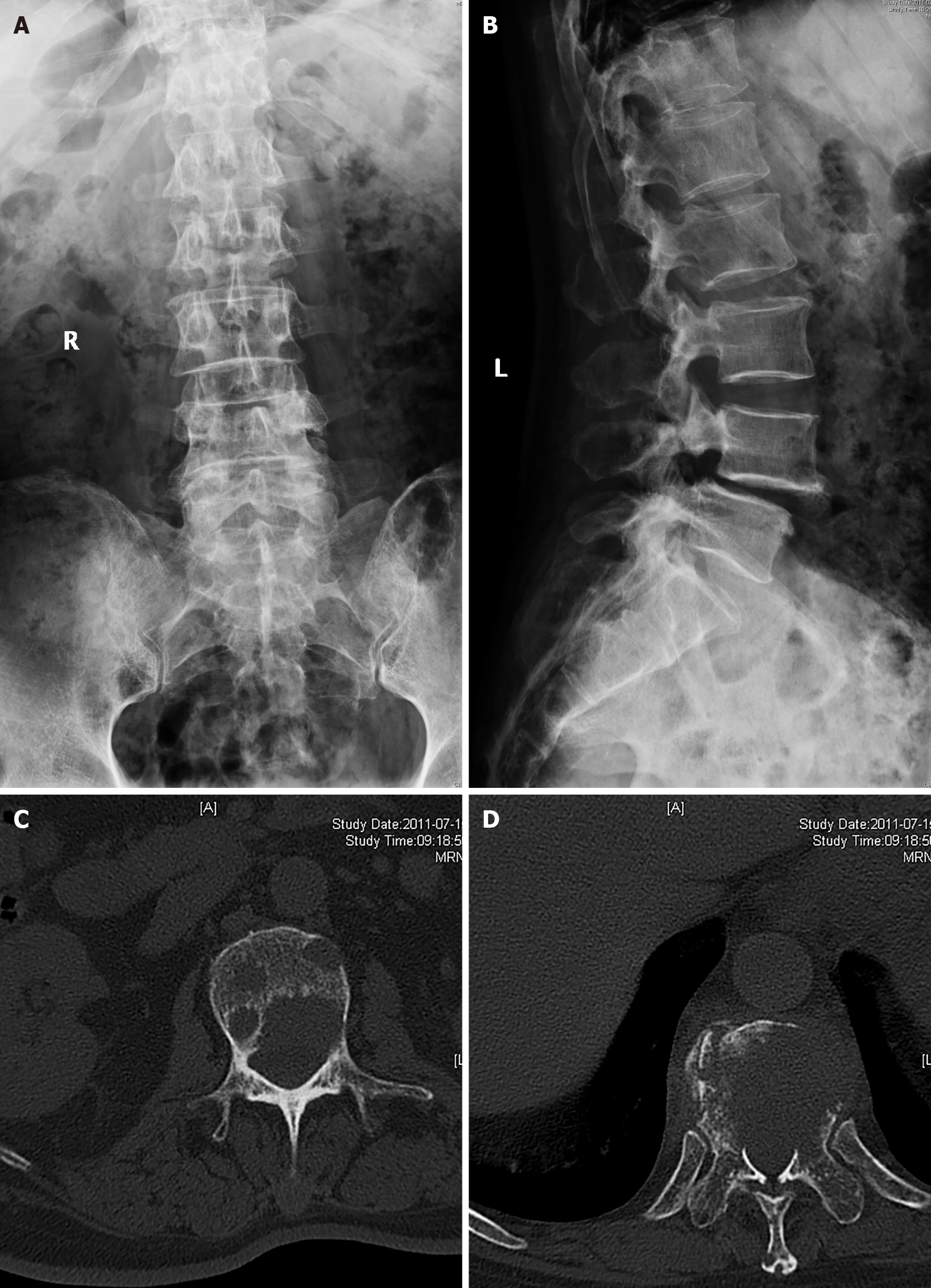
Figure 3 The score indicated “spinal instability”.
A, B: Lumbar spondylolisthesis is visible from the lateral view of the patient; Lumbar computed tomography cross-sectional image that shows an L2 lesion (C) and a T11 lesion (D).
- Citation: Yao XC, Shi XJ, Xu ZY, Tan J, Wei YZ, Qi L, Zhou ZH, Du XR. Preliminary establishment of a spinal stability scoring system for multiple myeloma. World J Clin Cases 2021; 9(30): 9023-9037
- URL: https://www.wjgnet.com/2307-8960/full/v9/i30/9023.htm
- DOI: https://dx.doi.org/10.12998/wjcc.v9.i30.9023


