Copyright
©The Author(s) 2021.
World J Clin Cases. Oct 16, 2021; 9(29): 8906-8914
Published online Oct 16, 2021. doi: 10.12998/wjcc.v9.i29.8906
Published online Oct 16, 2021. doi: 10.12998/wjcc.v9.i29.8906
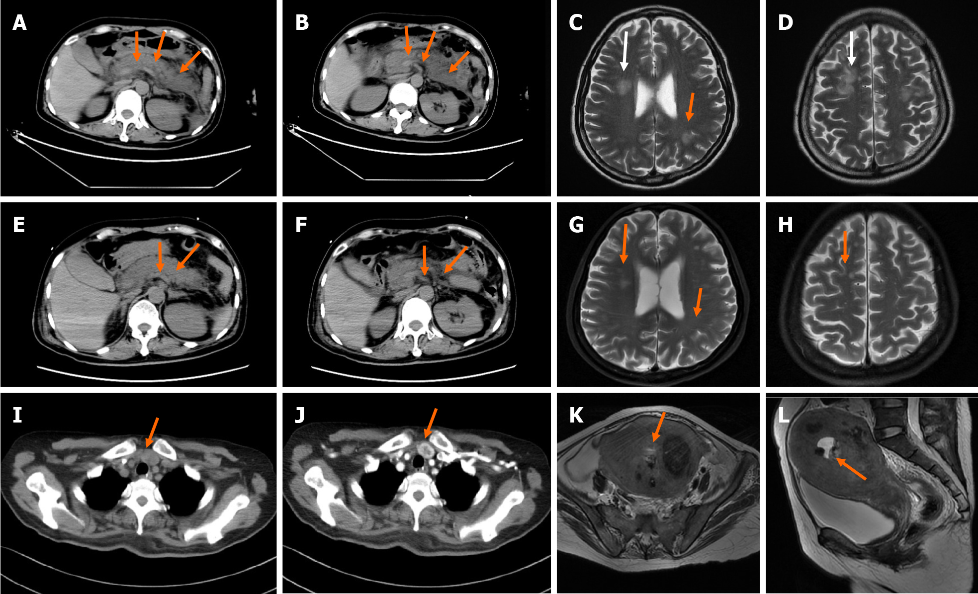
Figure 1 Computed tomography.
A and B: A non-contrast enhanced computed tomography (CT) scan of the abdomen revealed exudation around the pancreas (arrow); C and D: Head magnetic resonance imaging (MRI) revealed abnormal signal in bilateral fronto-parietal-temporal-occipital cortex-medullary junction area and bilateral paraventricular; E-H: A repeated non-contrast enhanced CT scan of the abdomen and head MRI showed reduction of pancreatic exudation and abnormal head signals after effective treatment; I and J: A contrast enhanced CT of the neck showed nodules located on the junction of left lobe of the thyroid and parathyroid gland; K and L: MRI of the pelvis suggested malignant lesions of the uterus and multiple uterine fibroids.
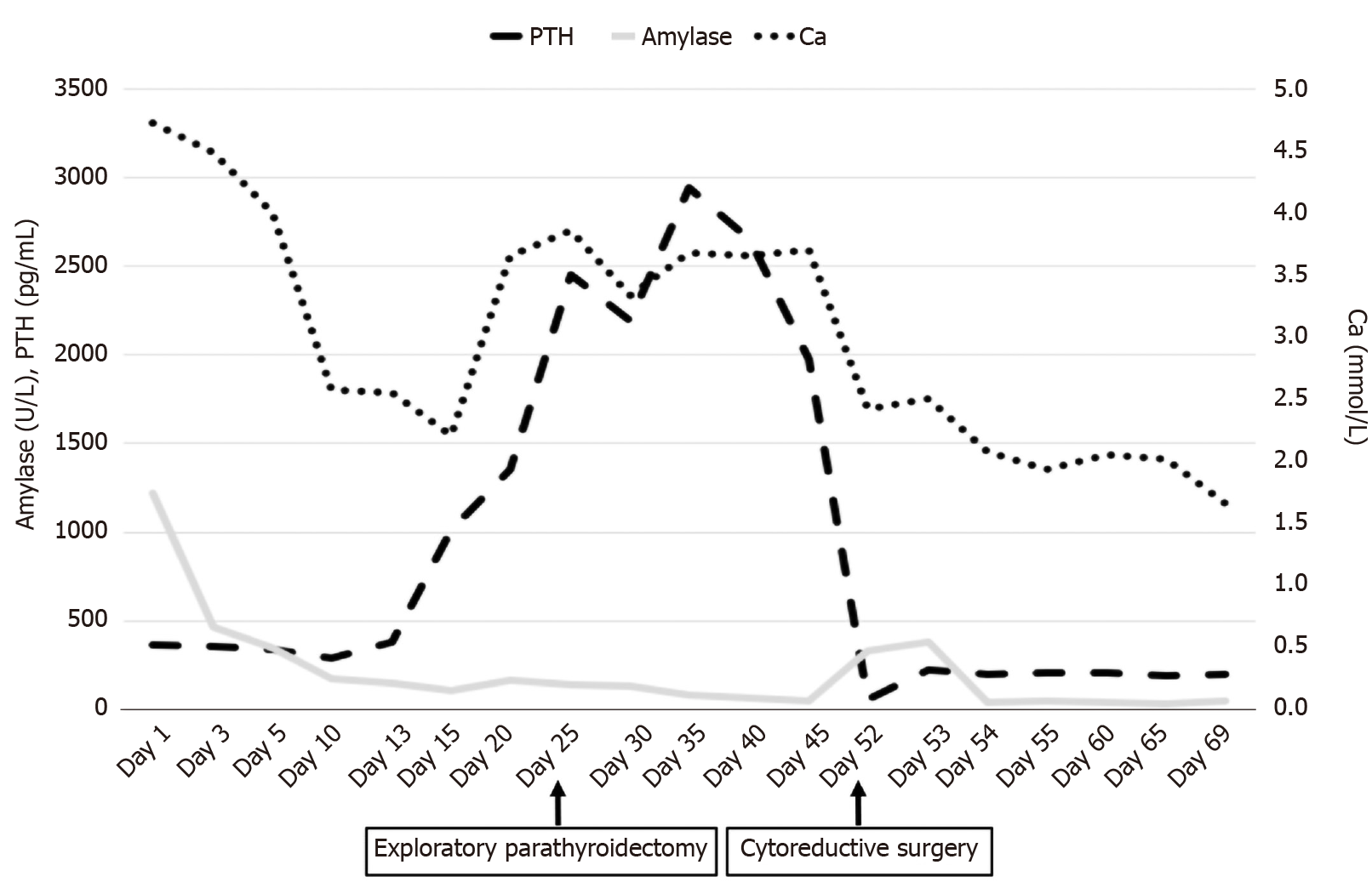
Figure 2 The serum concentration of amylase, calcium, and intact parathyroid hormone during the disease course of this patient.
The serum amylase gradually recovered to normal level whereas the intact parathyroid hormone concentration decreased after exploratory parathyroidectomy, which was performed 25 d after admission. Refractory hypercalcemia ultimately decreased to normal level after cytoreductive surgery on May 11. PTH: Parathyroid hormone.
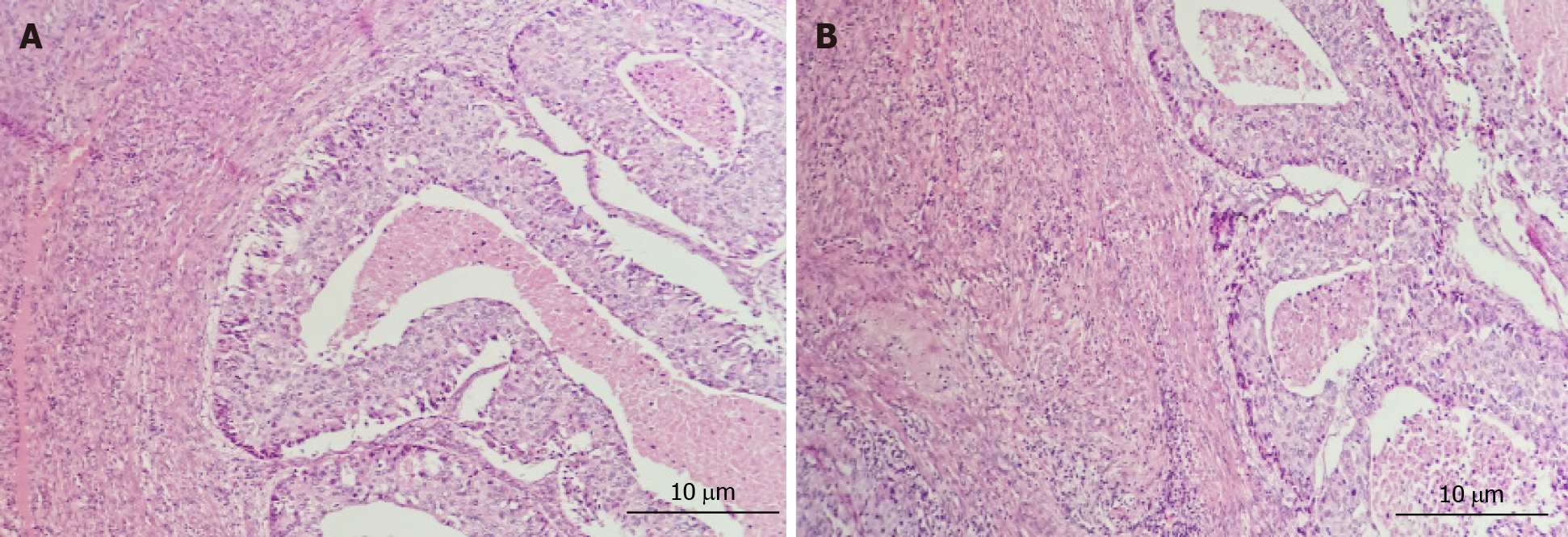
Figure 3 Pathological diagrams of endometrium.
Hematoxylin and eosin stain, × 200 magnification. A: Poorly differentiated endometrial carcinoma, increased lymphocytes infiltrate the cervical endometrium; B: Endometrioid carcinoma with squamous differentiation is not excepted, cytoplasm with eosinophilic keratinocytes, endometrial glandular differentiation.
- Citation: Yang L, Lin Y, Zhang XQ, Liu B, Wang JY. Acute pancreatitis with hypercalcemia caused by primary hyperparathyroidism associated with paraneoplastic syndrome: A case report and review of literature. World J Clin Cases 2021; 9(29): 8906-8914
- URL: https://www.wjgnet.com/2307-8960/full/v9/i29/8906.htm
- DOI: https://dx.doi.org/10.12998/wjcc.v9.i29.8906


