Copyright
©The Author(s) 2021.
World J Clin Cases. Oct 16, 2021; 9(29): 8782-8788
Published online Oct 16, 2021. doi: 10.12998/wjcc.v9.i29.8782
Published online Oct 16, 2021. doi: 10.12998/wjcc.v9.i29.8782
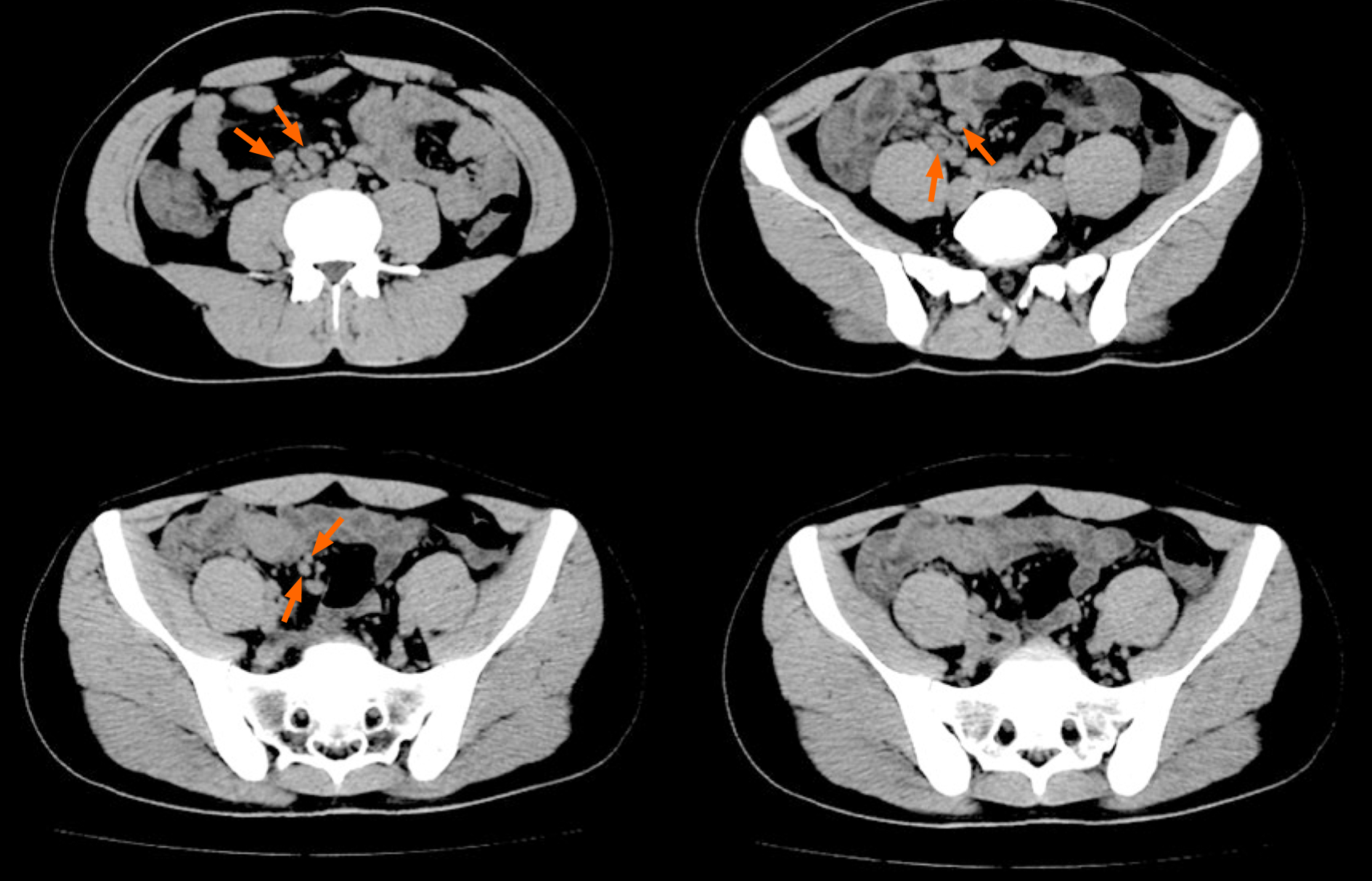
Figure 1 Pre-operative abdominal computed tomography showed a thickened intestinal wall of the ileocecal junction with multiple enlarged lymph nodes (orange arrows) nearby.
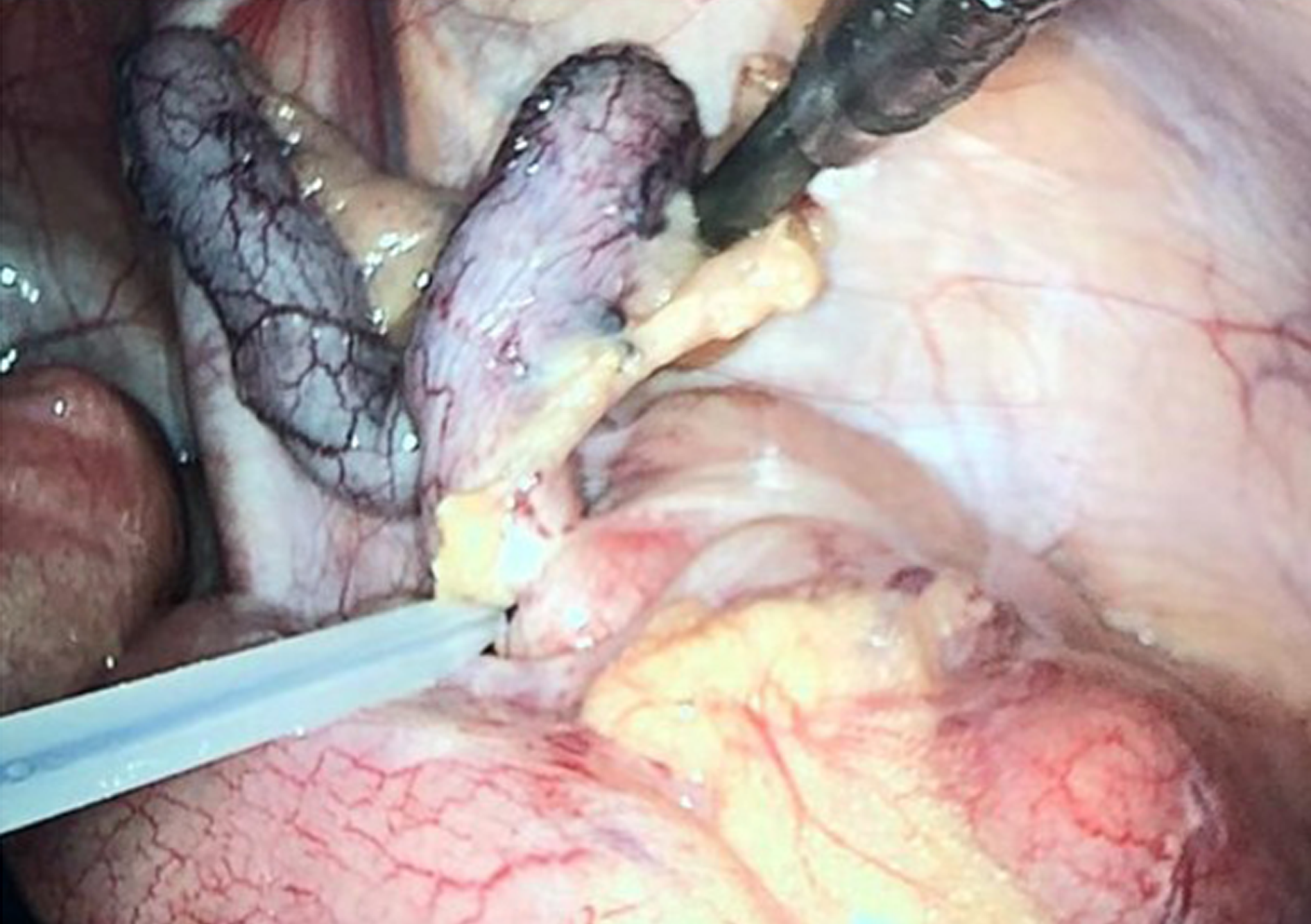
Figure 2 Intraoperative image of the appendix.
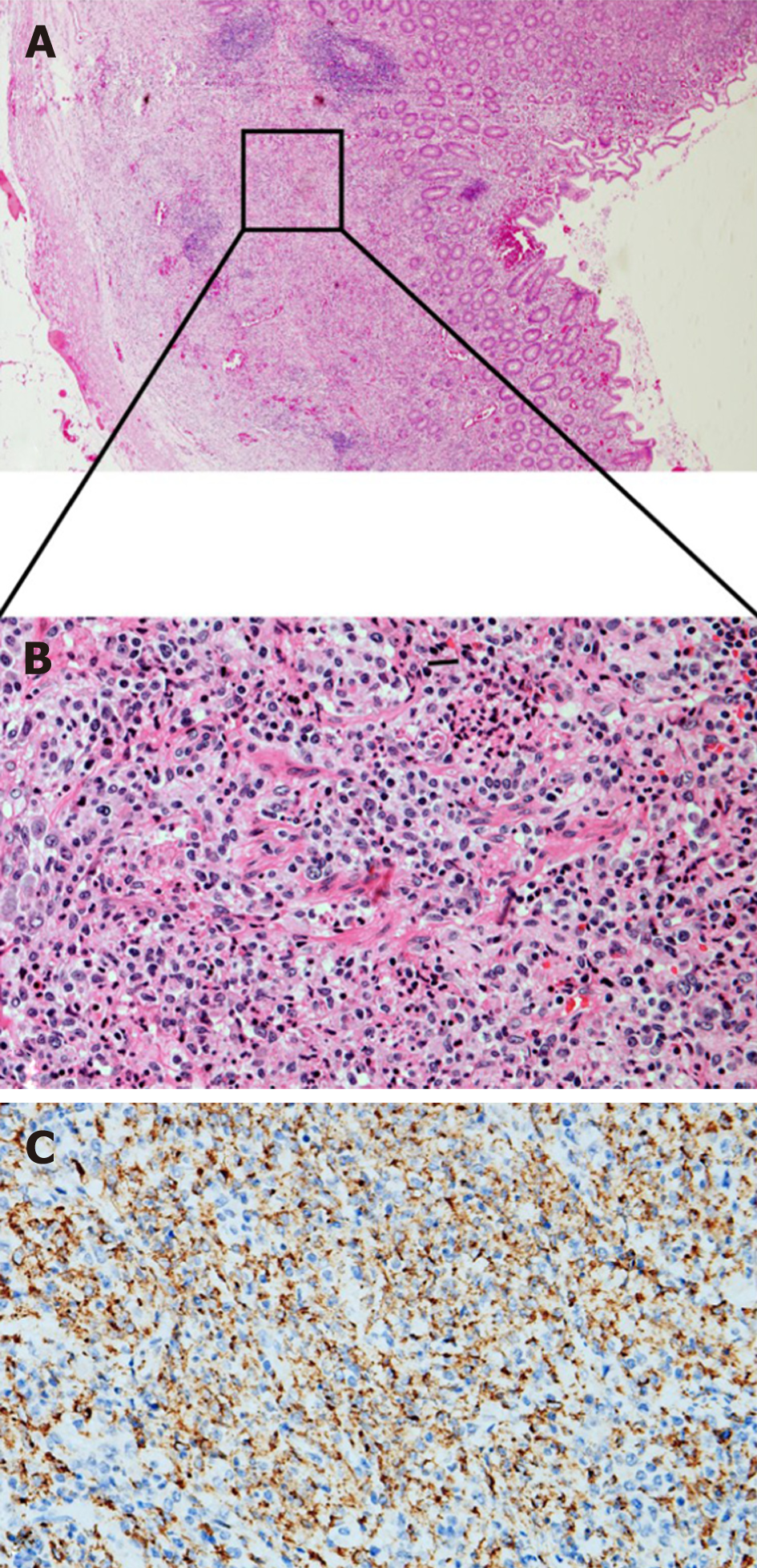
Figure 3 Pathological images of Samonellatyphi infection-related appendicitis.
A: Macrophage reactive hyperplasia in the submucosa is visible, and the normal lymphoid follicular structure disappears in the lamina propria (Hematoxylin-eosin staining, original magnification × 40); B: A magnification scope of Figure A, which shows massive macrophage reactive hyperplasia with a small amount of neutrophil and lymphocyte infiltration (Hematoxylin-eosin staining, original magnification × 400); C: Immunohistochemical staining for CD68 (Original magnification × 400).
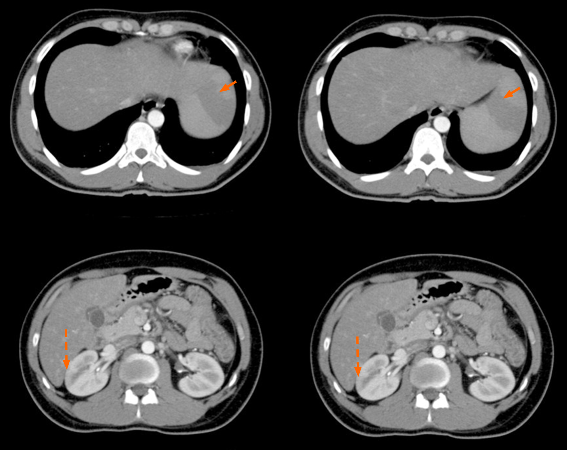
Figure 4 Post-operative abdominal computed tomography showed swelling of the cecal and ascending colonic wall, with multiple enlarged celiac and retroperitoneal lymph nodes, multiple spleen infarctions (orange arrows in solid line), and right renal infarction (orange arrows in dotted line).
- Citation: Zheng BH, Hao WM, Lin HC, Shang GG, Liu H, Ni XJ. Samonella typhi infection-related appendicitis: A case report. World J Clin Cases 2021; 9(29): 8782-8788
- URL: https://www.wjgnet.com/2307-8960/full/v9/i29/8782.htm
- DOI: https://dx.doi.org/10.12998/wjcc.v9.i29.8782


