Copyright
©The Author(s) 2021.
World J Clin Cases. Sep 26, 2021; 9(27): 8177-8185
Published online Sep 26, 2021. doi: 10.12998/wjcc.v9.i27.8177
Published online Sep 26, 2021. doi: 10.12998/wjcc.v9.i27.8177
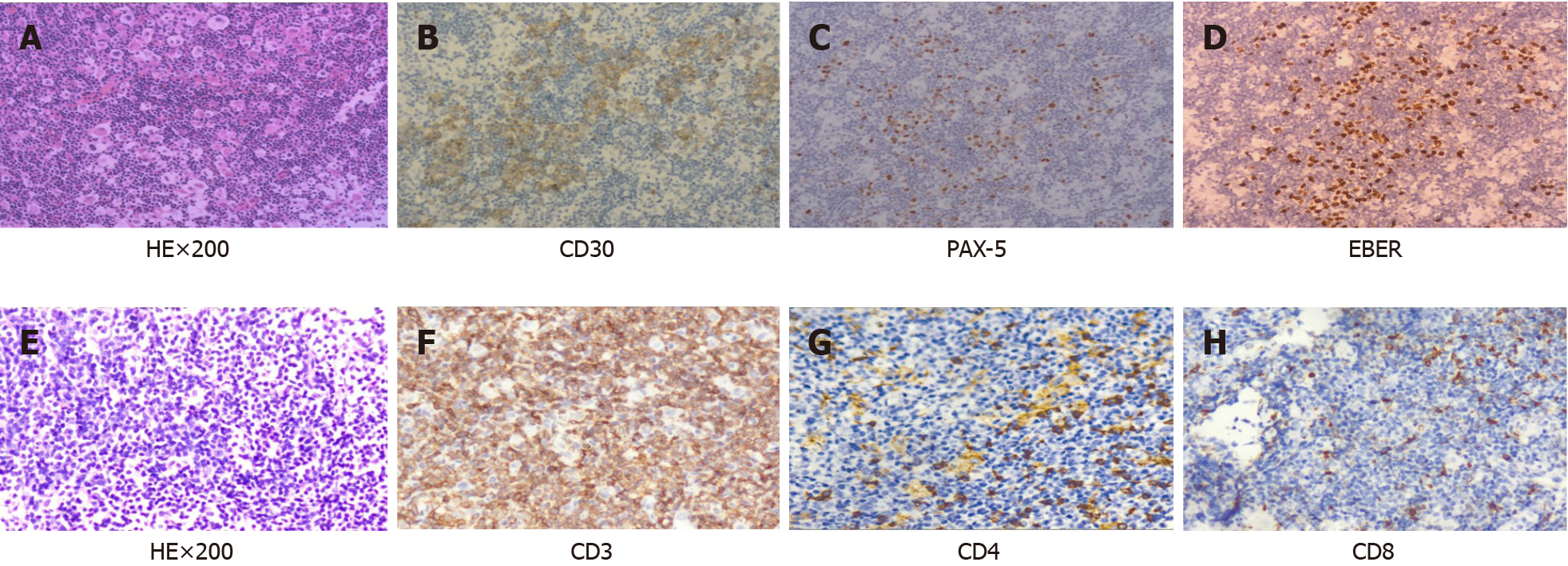
Figure 1 Pathological findings of Hodgkin’s lymphoma and the peripheral T-cell lymphoma.
A-D: Pathological findings of Hodgkin’s lymphoma; the lymph node structure was disrupted, and a large number of small lymphocytes and popcorn-like cells were observed, with abundant blood vessels (HE, × 200) (A), positive expression of CD30 (B) and PAX-5 (C) in reed-sternberg (RS) cells, EBER+ expression in RS cells (D); E-H: Pathological findings of the peripheral T-cell lymphoma; the lymph node structure was disrupted, and diffuse consistent T-cells were observed, with abundant blood vessels (HE, × 200) (E), CD3+ T-cells (F), CD4+ (G) and CD8+ T-cells (few) (H).
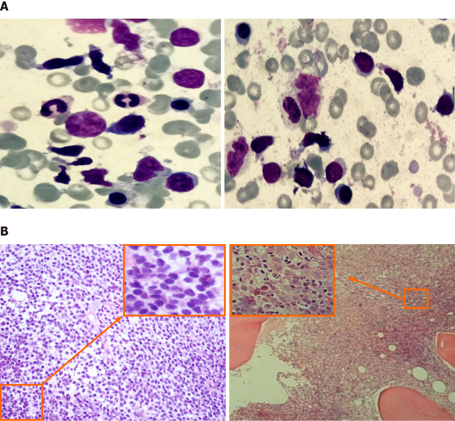
Figure 2 Morphological and biopsy results of peripheral T-cell lymphoma bone marrow.
A: Atypical lymphocytes, with irregular, spindle-shaped, small to medium morphology, visible pseudopods and cytoplasmic granules, and no reed-sternberg cells observed (× 100); B: The lymphoma cells were diffusely proliferating, with small cytosol, low cytoplasmic volume, irregular nuclei, and coarse chromatin (HE/periodic acid-schiff, × 40 and × 400).
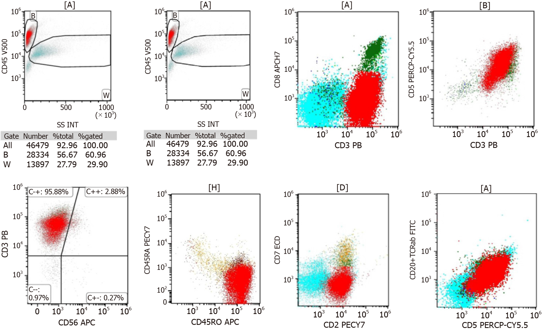
Figure 3 Immunophenotyping of T/natural killer lymphoma through 8-color flow cytometry.
Abnormal T lymphocytes accounted for 57.8% of the nucleated cells and expressed CD3+, CD2+, CD5+, TCRαβ, and CD45RO.
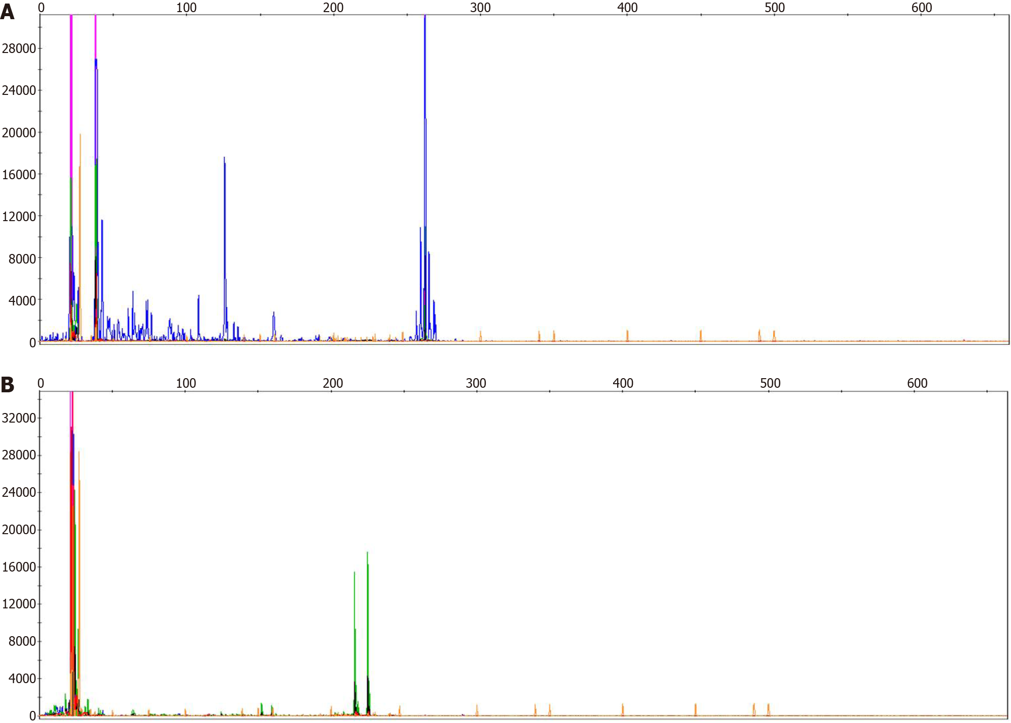
Figure 4 Identification of the T-cell receptor clone gene rearrangement by applying BIOMED-2 primer system.
A: TCRβ gene rearrangement were detected; B: TCRγ gene rearrangement were detected.
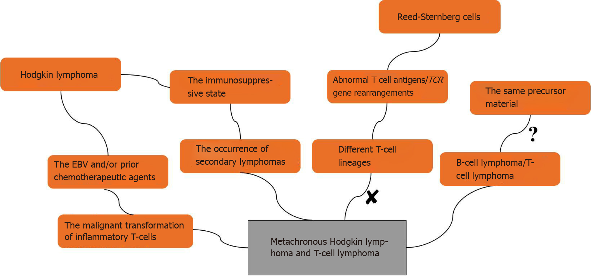
Figure 5 The possible mechanisms of metachronous B-cell lymphoma and T-cell lymphoma.
EBV: Epstein-Barr virus.
- Citation: Dong Y, Deng LJ, Li MM. Metachronous mixed cellularity classical Hodgkin’s lymphoma and T-cell leukemia/lymphoma: A case report. World J Clin Cases 2021; 9(27): 8177-8185
- URL: https://www.wjgnet.com/2307-8960/full/v9/i27/8177.htm
- DOI: https://dx.doi.org/10.12998/wjcc.v9.i27.8177


