Copyright
©The Author(s) 2021.
World J Clin Cases. Sep 26, 2021; 9(27): 8135-8141
Published online Sep 26, 2021. doi: 10.12998/wjcc.v9.i27.8135
Published online Sep 26, 2021. doi: 10.12998/wjcc.v9.i27.8135
Figure 1 Abdominal computerized tomography scans show continuous increase of the calcifications following the initiation of chemotherapy.
A: May 2020; B: September 2020; C: December 2020; D: March 2021.
Figure 2 Initial endoscopic examination.
A: Gastric body; B: Gastric lesser curvature.
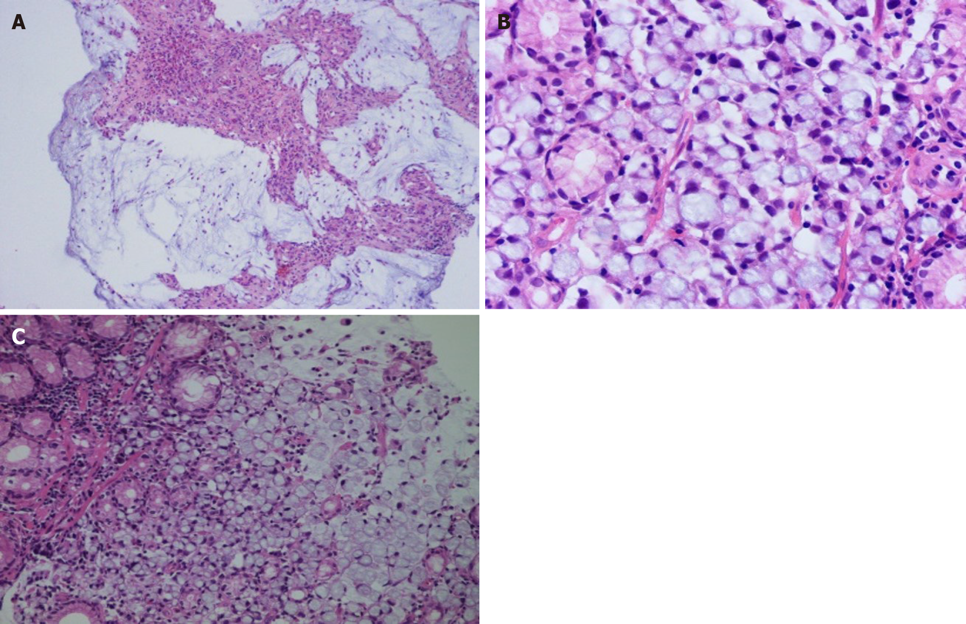
Figure 3 Histopathological examination shows mucinous gastric carcinoma with signet-ring cells.
A: Magnification: 100 ×; B: Magnification: 200 ×; C: Magnification: 400 ×.
- Citation: Lin YH, Yao W, Fei Q, Wang Y. Gastric cancer with calcifications: A case report. World J Clin Cases 2021; 9(27): 8135-8141
- URL: https://www.wjgnet.com/2307-8960/full/v9/i27/8135.htm
- DOI: https://dx.doi.org/10.12998/wjcc.v9.i27.8135


