Copyright
©The Author(s) 2021.
World J Clin Cases. Aug 16, 2021; 9(23): 6943-6949
Published online Aug 16, 2021. doi: 10.12998/wjcc.v9.i23.6943
Published online Aug 16, 2021. doi: 10.12998/wjcc.v9.i23.6943
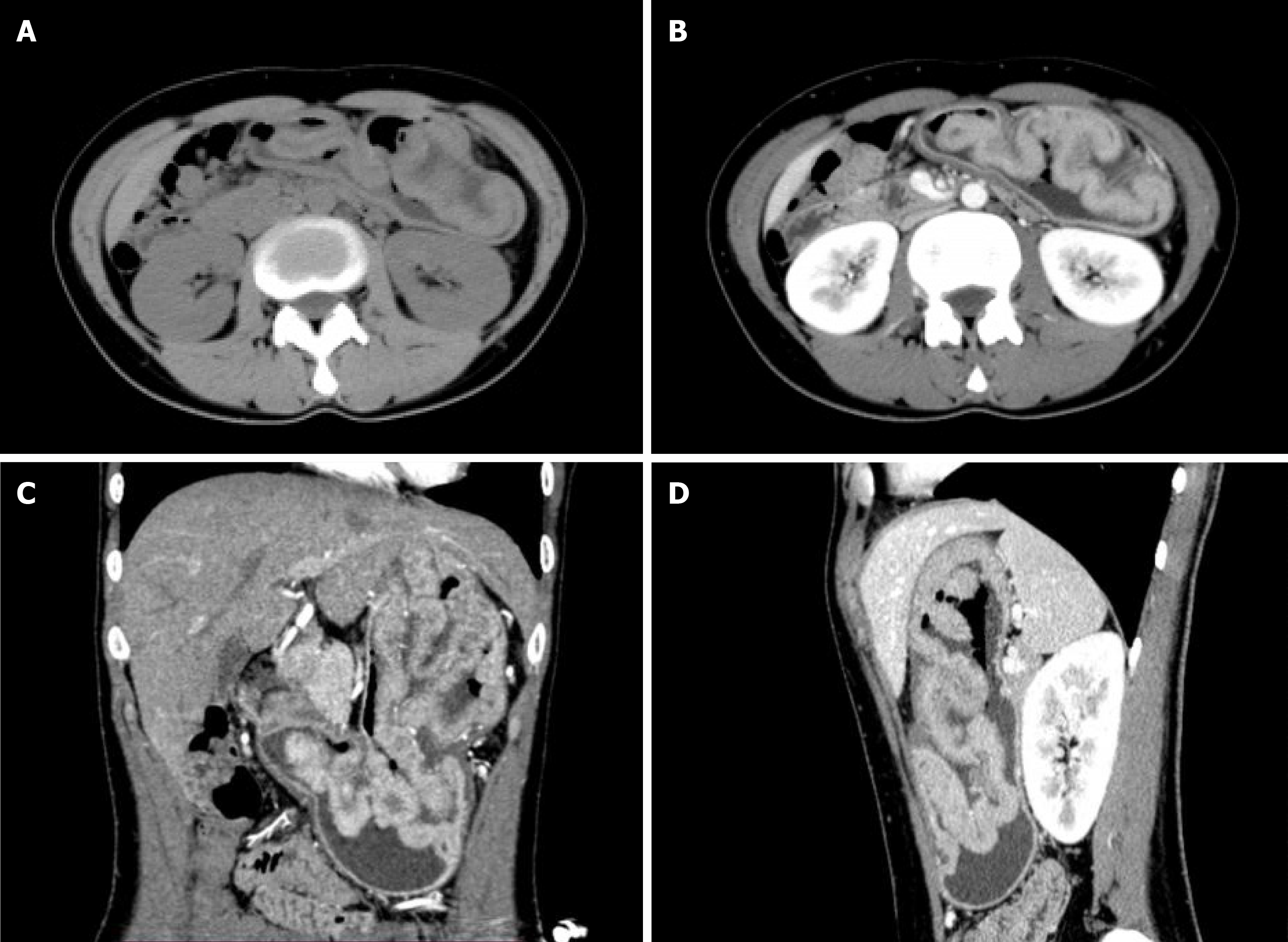
Figure 1 Abdominal computed tomography images.
A: Plain scan cross section; B: Enhanced scan cross section; C: Plain scan coronal plane; D: Enhanced scan coronal plane.
Figure 2 Abdominal magnetic resonance.
A: Flat scan cross section; B: Enhanced cross section; C: Enhanced coronal plane.
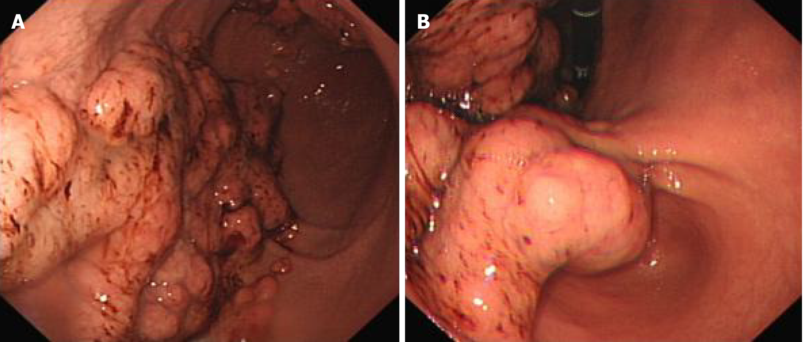
Figure 3 Gastrointestinal endoscopy images.
A: Irregular mucosal bulge at the bottom of the stomach; B: Irregular mucosal bulge in gastric antrum.
Figure 4 Histological images.
A: Hematoxylin-Eosin staining (×100); B: Hematoxylin-Eosin staining (× 400).
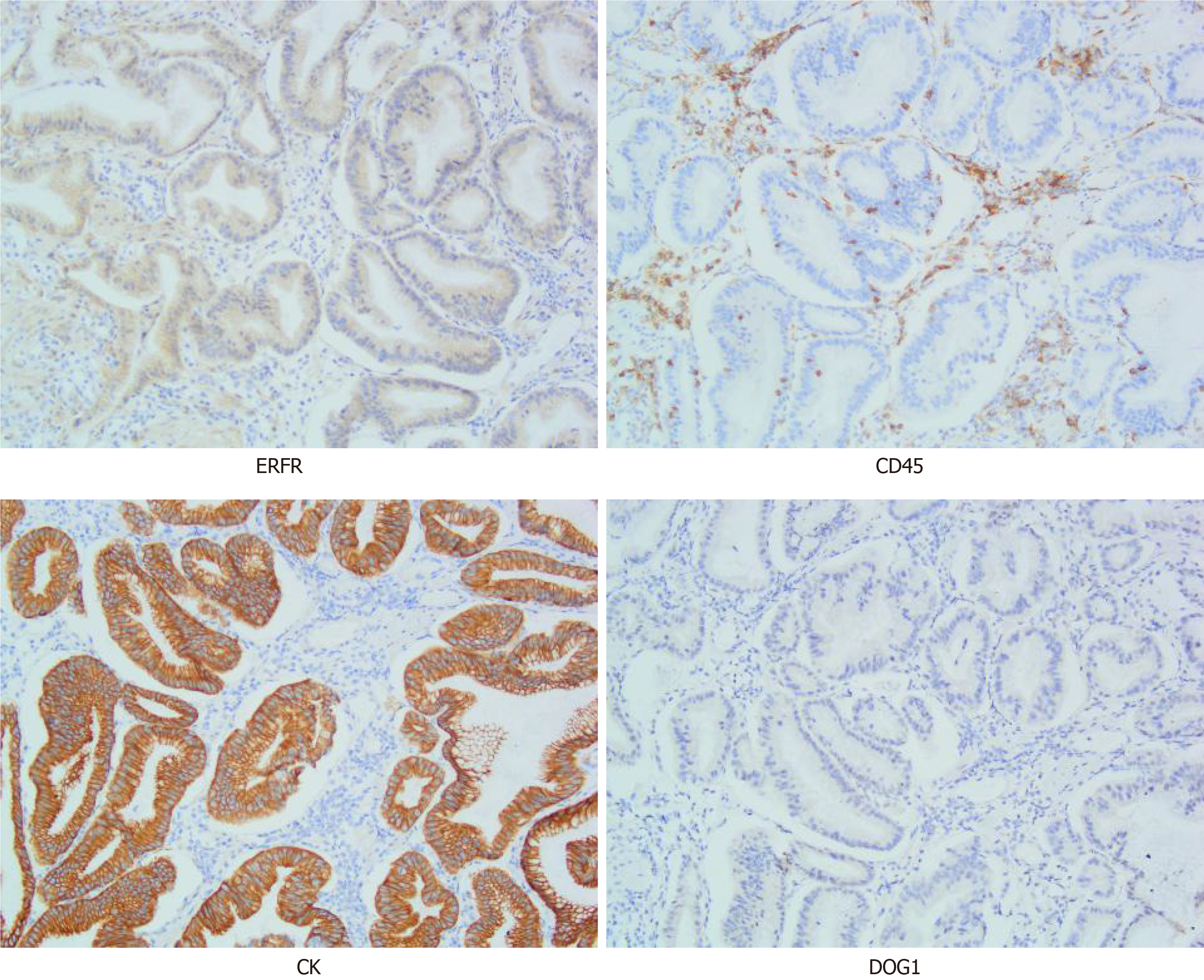
Figure 5 Immunohistochemical.
ERFR: Positive expression; CD45: Positive lymphocytes scattered among hyperplastic glands; CK: The hyperplastic glands scattered regularly; DOG1: Proliferating spindle cells between hyperplastic glands.
- Citation: Wang HH, Zhao CC, Wang XL, Cheng ZN, Xie ZY. Menetrier’s disease and differential diagnosis: A case report. World J Clin Cases 2021; 9(23): 6943-6949
- URL: https://www.wjgnet.com/2307-8960/full/v9/i23/6943.htm
- DOI: https://dx.doi.org/10.12998/wjcc.v9.i23.6943


