Copyright
©The Author(s) 2021.
World J Clin Cases. Aug 16, 2021; 9(23): 6907-6915
Published online Aug 16, 2021. doi: 10.12998/wjcc.v9.i23.6907
Published online Aug 16, 2021. doi: 10.12998/wjcc.v9.i23.6907
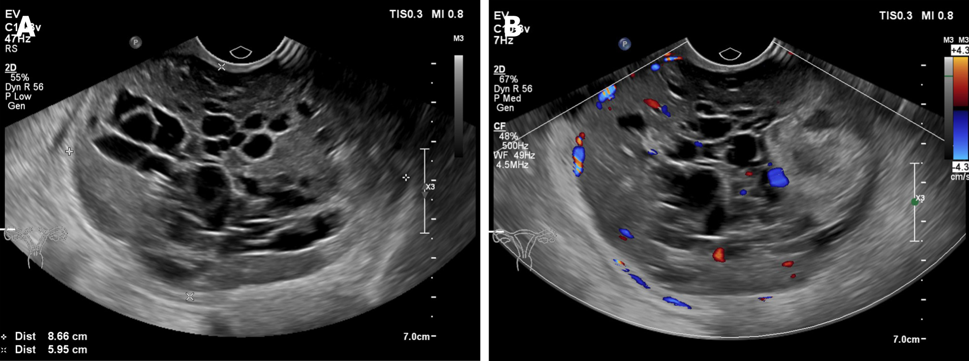
Figure 1 Ultrasound scan.
A: An 87 mm × 60 mm mass with a heterogeneous echo is seen on the right side of the pelvic cavity. It consists of a hypoechoic intracystic effusion and a hypoechoic intracystic tumor, with local honeycomb changes; B: Color Doppler imaging shows hypervascularity.
Figure 2 Contrast-enhanced computed tomography.
A: A round low-density mass with uneven internal density was present in the pelvic cavity; B, C: Uneven enhancement was present in the arterial and venous phases of the scan and no enhancement in low-density areas.
Figure 3 Histopathological findings.
A: Hematoxylin-eosin (HE) staining (× 100); B: HE staining (× 200); C: HE staining (× 400).
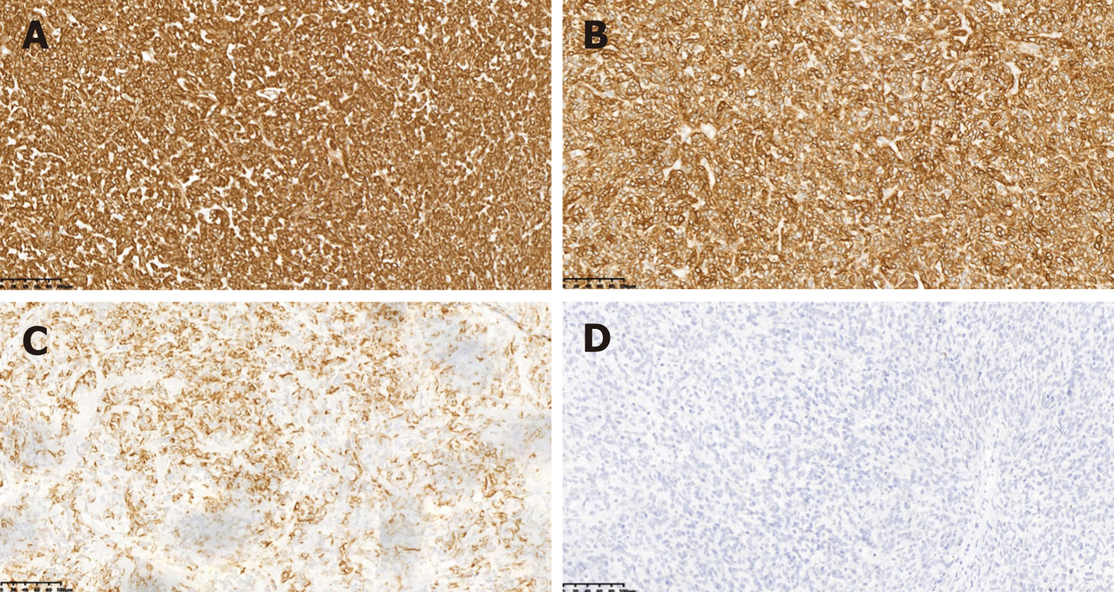
Figure 4 Immunohistochemical staining (× 100).
A: Immunostaining for vimentin; B: CD99; C: Cytokeratin; D: α-Inhibin.
- Citation: Zhou FF, He YT, Li Y, Zhang M, Chen FH. Uterine tumor resembling an ovarian sex cord tumor: A case report and review of literature. World J Clin Cases 2021; 9(23): 6907-6915
- URL: https://www.wjgnet.com/2307-8960/full/v9/i23/6907.htm
- DOI: https://dx.doi.org/10.12998/wjcc.v9.i23.6907


