Copyright
©The Author(s) 2021.
World J Clin Cases. Aug 6, 2021; 9(22): 6388-6392
Published online Aug 6, 2021. doi: 10.12998/wjcc.v9.i22.6388
Published online Aug 6, 2021. doi: 10.12998/wjcc.v9.i22.6388
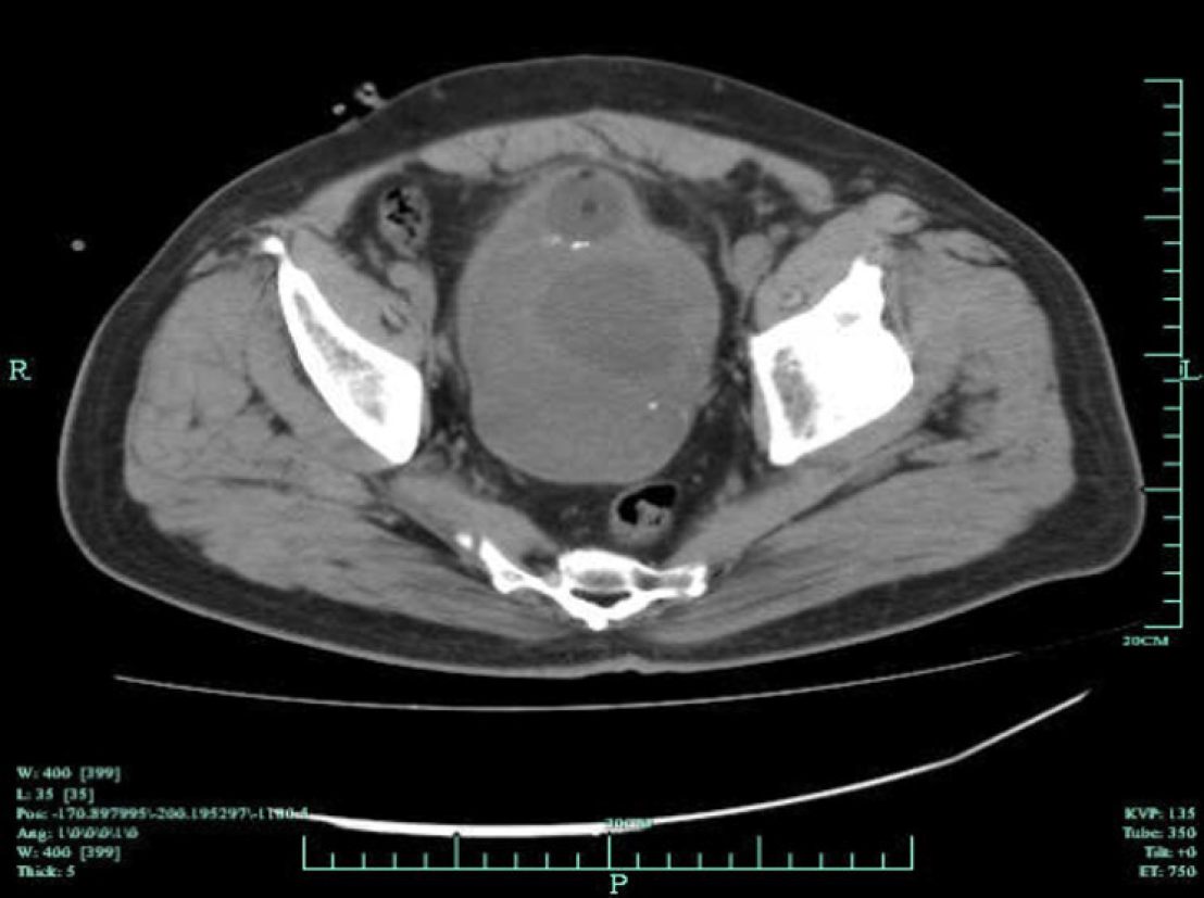
Figure 1 Preoperative pelvic computed tomography image demonstrating a 15 cm × 9 cm × 8 cm-large tumor mass with central necrosis distorting the bladder neck, which could only be recognized by a catheter balloon.
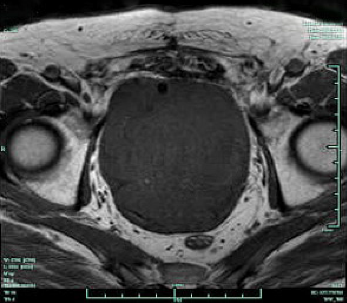
Figure 2
Preoperative pelvic magnetic resonance image showing a large tumor mass occupying the majority of the pelvic cavity with no evidence of rectal metastasis.
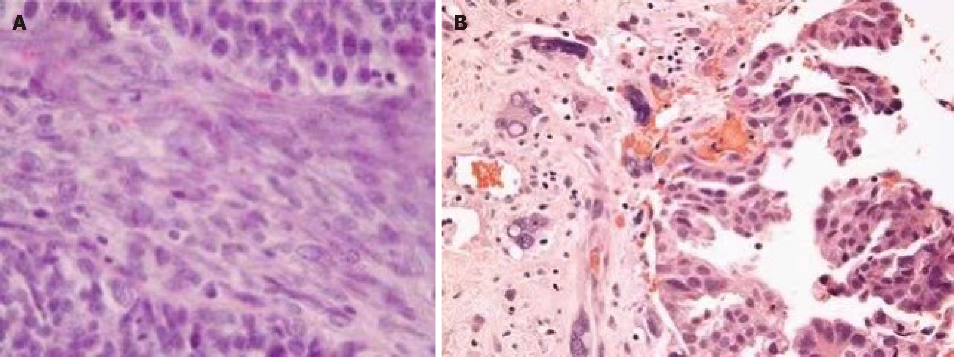
Figure 3 Hematoxylin–eosin staining.
A: Image showing the pelvic tumor with sarcomatous-component spindled cells (original magnification, × 400); B: Image showing the pelvic tumor with sarcomatous-bizarre cells (original magnification, × 400).
- Citation: Huang X, Cai SL, Xie LP. Prostatic carcinosarcoma seven years after radical prostatectomy and hormonal therapy for prostatic adenocarcinoma: A case report. World J Clin Cases 2021; 9(22): 6388-6392
- URL: https://www.wjgnet.com/2307-8960/full/v9/i22/6388.htm
- DOI: https://dx.doi.org/10.12998/wjcc.v9.i22.6388


