Copyright
©The Author(s) 2021.
World J Clin Cases. Aug 6, 2021; 9(22): 6244-6253
Published online Aug 6, 2021. doi: 10.12998/wjcc.v9.i22.6244
Published online Aug 6, 2021. doi: 10.12998/wjcc.v9.i22.6244
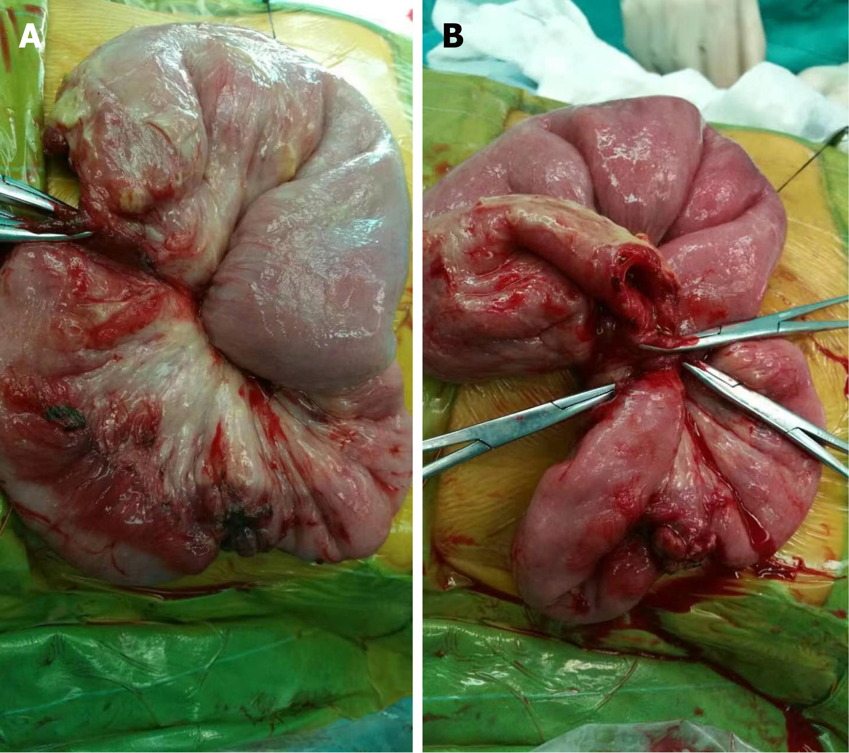
Figure 1 Image of the small intestine of a 6-year-old girl who had intussusception and intestinal necrosis.
A and B: A 6-year-old girl presented with a 12-h history of abdominal pain, vomiting, and purpura. She was diagnosed with Henoch-Schönlein purpura and intussusception. During the operation, the head of intussusception was found in the jejunum 50 cm away from the torus ligament, and the tail was found in the small intestine 30 cm away from the torus ligament. After reduction, the intestinal tubes were found to be black and purple in color. Enterectomy and anastomosis were performed, and the length of the diseased intestinal tubes was about 30 cm.
Figure 2 Pathological images of resected intestinal tissue.
A: The intestinal tissue had hyperemia, bleeding, mucosal epithelial necrosis, ulcer formation, granulation tissue hyperplasia, and high submucosal edema; B: The vascular wall was slightly thickened, and some of the vascular wall structure was destroyed. Flake-like infiltration of neutrophils and lymphocytes was observed in the wall and around the small vessel, and many nuclear fragments were observed.
- Citation: Zhao Q, Yang Y, He SW, Wang XT, Liu C. Risk factors for intussusception in children with Henoch-Schönlein purpura: A case-control study. World J Clin Cases 2021; 9(22): 6244-6253
- URL: https://www.wjgnet.com/2307-8960/full/v9/i22/6244.htm
- DOI: https://dx.doi.org/10.12998/wjcc.v9.i22.6244


