Copyright
©The Author(s) 2021.
World J Clin Cases. Jul 16, 2021; 9(20): 5621-5630
Published online Jul 16, 2021. doi: 10.12998/wjcc.v9.i20.5621
Published online Jul 16, 2021. doi: 10.12998/wjcc.v9.i20.5621
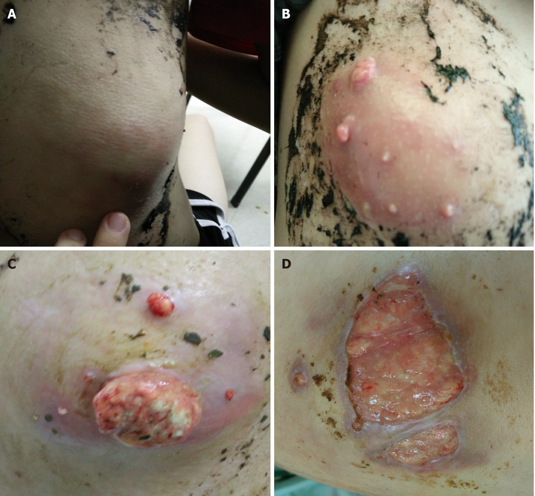
Figure 1
These images show the successive changes (from A to D) of the skin lesion on the lower right back over several months up to September 30, 2018.
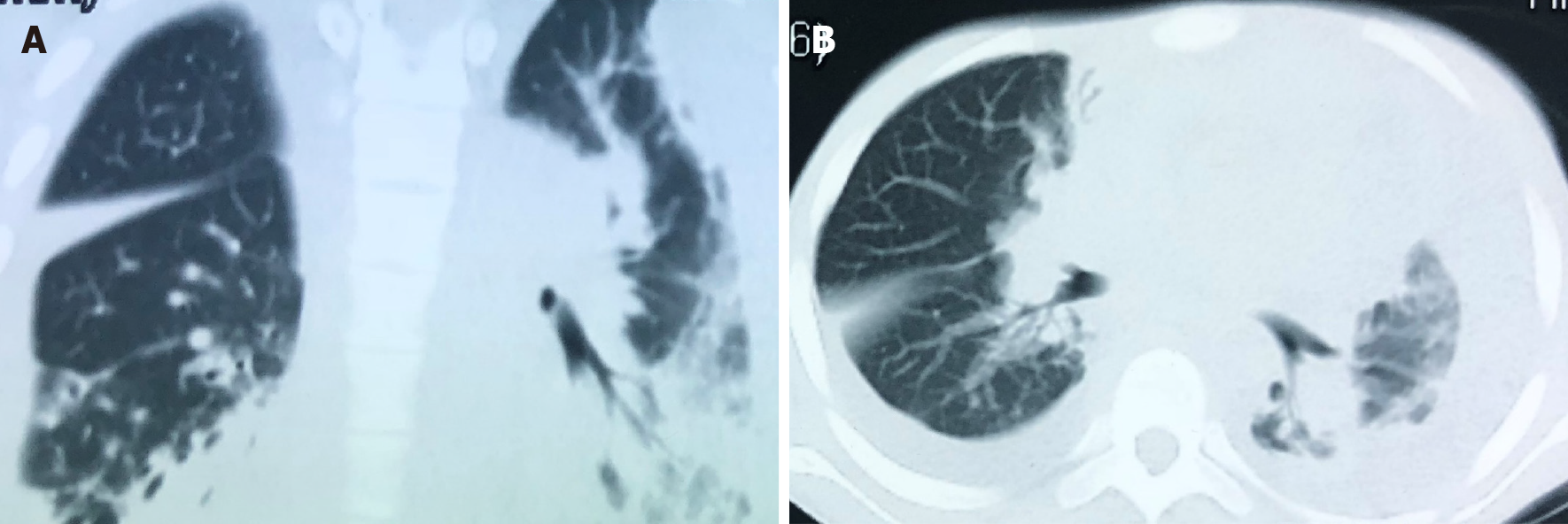
Figure 2 The chest computed tomography on September 21, 2018 shows a massive pleural effusion on the left, a small pleural effusion on the right, patchy and streaky cloudy opacities on both lungs, suggesting inflammation.
A: The sagittal plane; B: The horizontal plane.
Figure 3 The pathological manifestations of abscess tissue on the lower right back shows numerous lymphocytes and neutrophils infiltration, a small number of multinuclear giant cells and granulation tissues were found, which confirmed suppurative and granulomatous inflammation.
A: Magnification of 200 ×, B: Magnification of 400 ×.
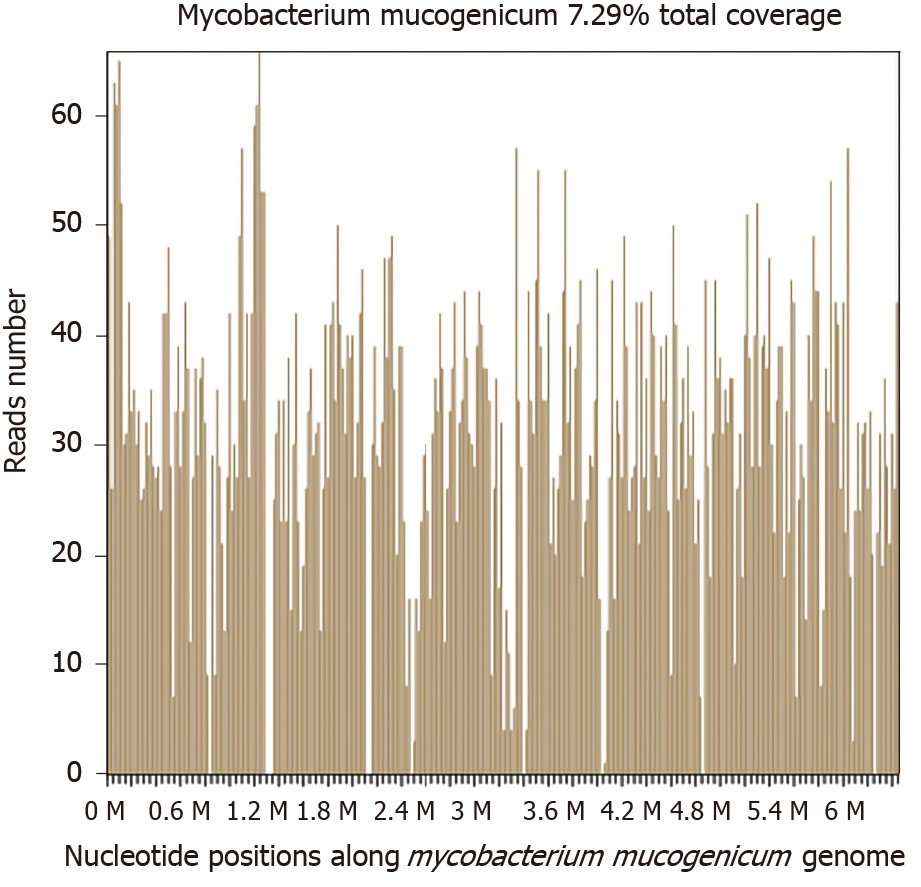
Figure 4
Sequencing results and phylogenetic analysis show nucleotide positions in the Mycobacteriummucogenicum genome.
Figure 5 These images show the four wounds on the 12th day after anti-nontuberculous mycobacteria treatment.
A: A mass on the right upper abdomen; B: A mass on the left lower back; C: Two masses on the right back.
Figure 6 These images show the four wounds on the 90th day after anti-nontuberculous mycobacteria treatment.
A: A mass on the right upper abdomen, B: A mass on the left lower back; C: Two masses on the right back.
Figure 7 The chest computed tomography on January 23, 2019 shows pleural effusion on both sides were absorbed completely, and patchy and streaky cloudy opacities on both lungs were improved compared with the images in Figure 2.
A: The sagittal plane; B: The horizontal plane.
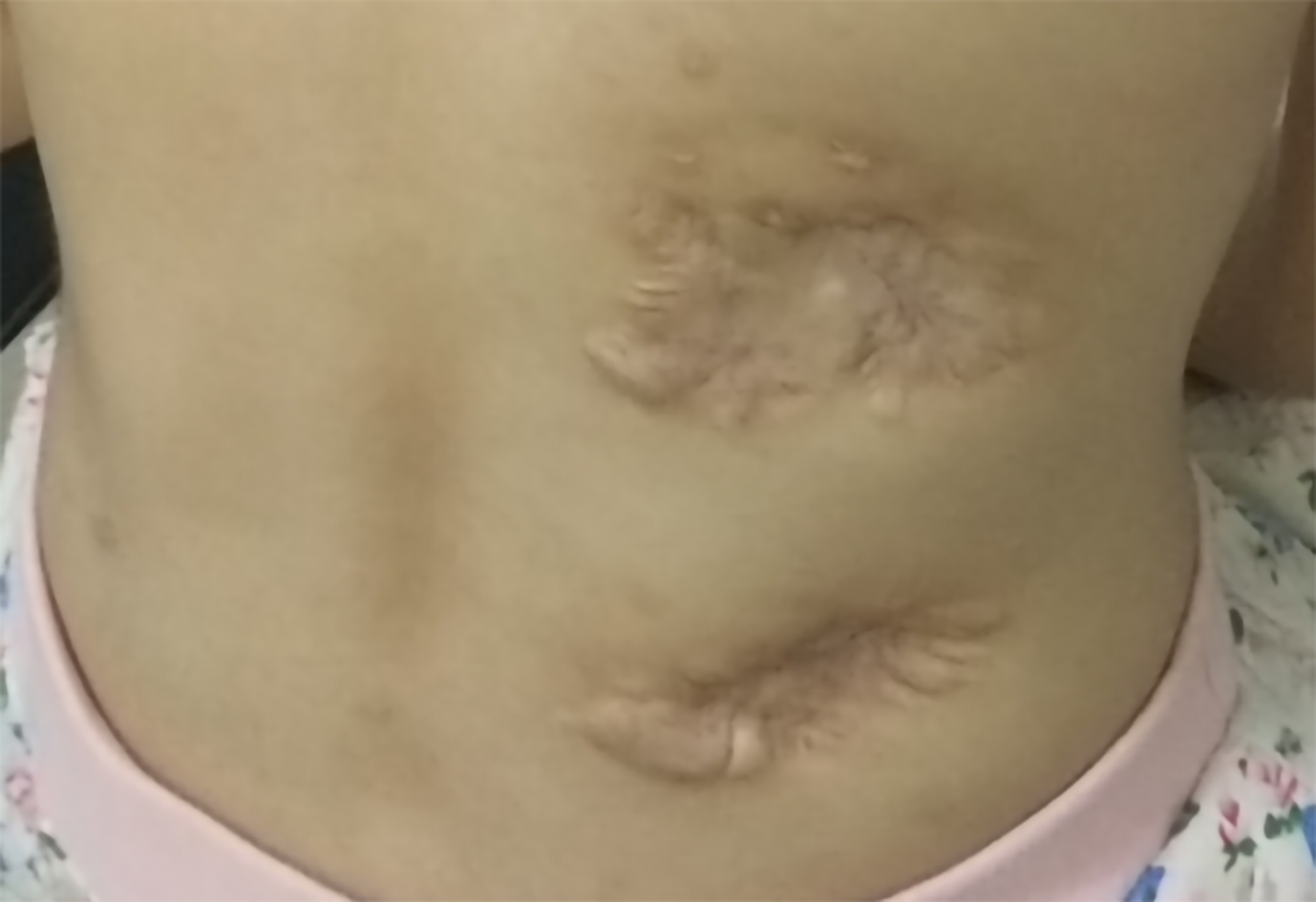
Figure 8
This image shows two masses on the right back in February, 2021.
- Citation: Liu J, Lei ZY, Pang YH, Huang YX, Xu LJ, Zhu JY, Zheng JX, Yang XH, Lin BL, Gao ZL, Zhuo C. Rapid diagnosis of disseminated Mycobacterium mucogenicum infection in formalin-fixed, paraffin-embedded specimen using next-generation sequencing: A case report. World J Clin Cases 2021; 9(20): 5621-5630
- URL: https://www.wjgnet.com/2307-8960/full/v9/i20/5621.htm
- DOI: https://dx.doi.org/10.12998/wjcc.v9.i20.5621


