Copyright
©The Author(s) 2021.
World J Clin Cases. Jul 16, 2021; 9(20): 5611-5620
Published online Jul 16, 2021. doi: 10.12998/wjcc.v9.i20.5611
Published online Jul 16, 2021. doi: 10.12998/wjcc.v9.i20.5611
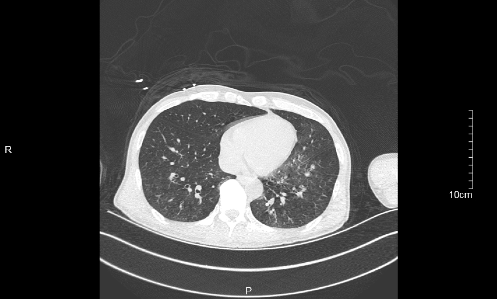
Figure 1 Emergency chest computed tomography.
Emergency chest computed tomography indicated bilateral pneumonia with bilateral pleural effusion.
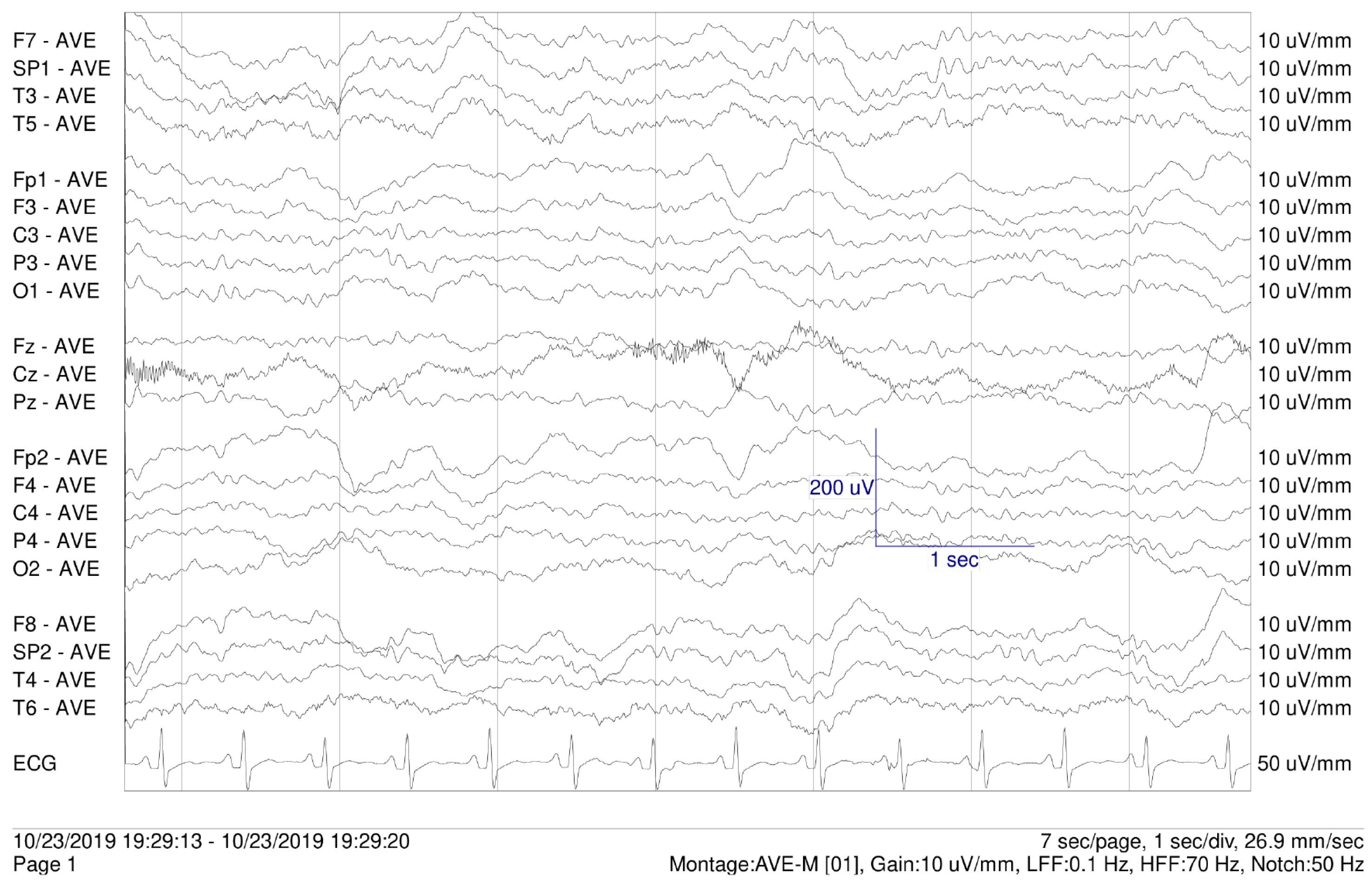
Figure 2 Electroencephalogram on the 3rd day.
Electroencephalogram showed slow-wave and no epileptiform discharges.
- Citation: Le DS, Su H, Liao ZL, Yu EY. Low-dose clozapine-related seizure: A case report and literature review . World J Clin Cases 2021; 9(20): 5611-5620
- URL: https://www.wjgnet.com/2307-8960/full/v9/i20/5611.htm
- DOI: https://dx.doi.org/10.12998/wjcc.v9.i20.5611


