Copyright
©The Author(s) 2021.
World J Clin Cases. Jul 16, 2021; 9(20): 5605-5610
Published online Jul 16, 2021. doi: 10.12998/wjcc.v9.i20.5605
Published online Jul 16, 2021. doi: 10.12998/wjcc.v9.i20.5605
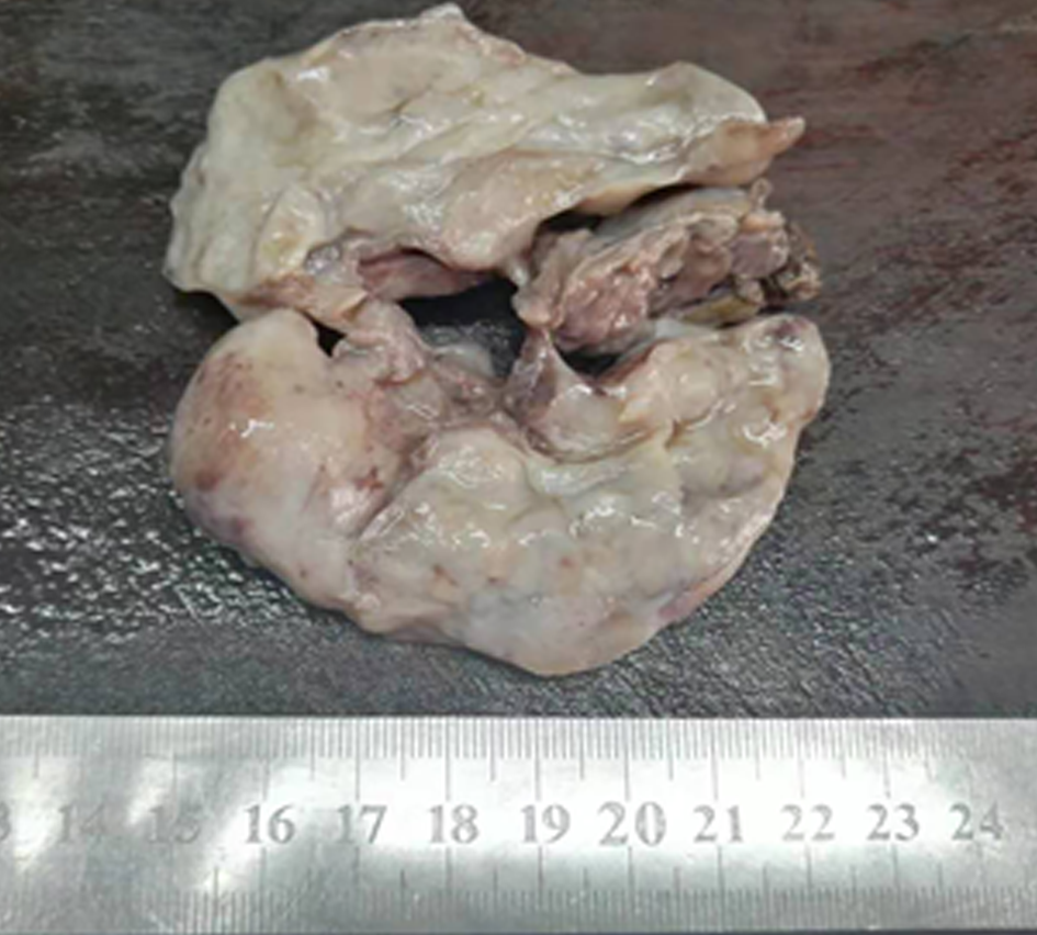
Figure 1 Postoperative gross pathology.
A subcutaneous tumor approximately 8 cm in diameter was observed in the perianal area.
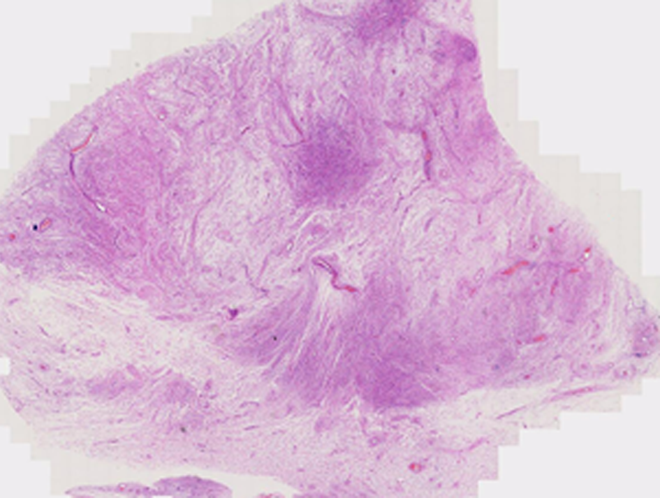
Figure 2 Histopathological examination by hematoxylin-eosin staining (0.
45 ×). A tumor with a clear boundary was located in the upper dermis.
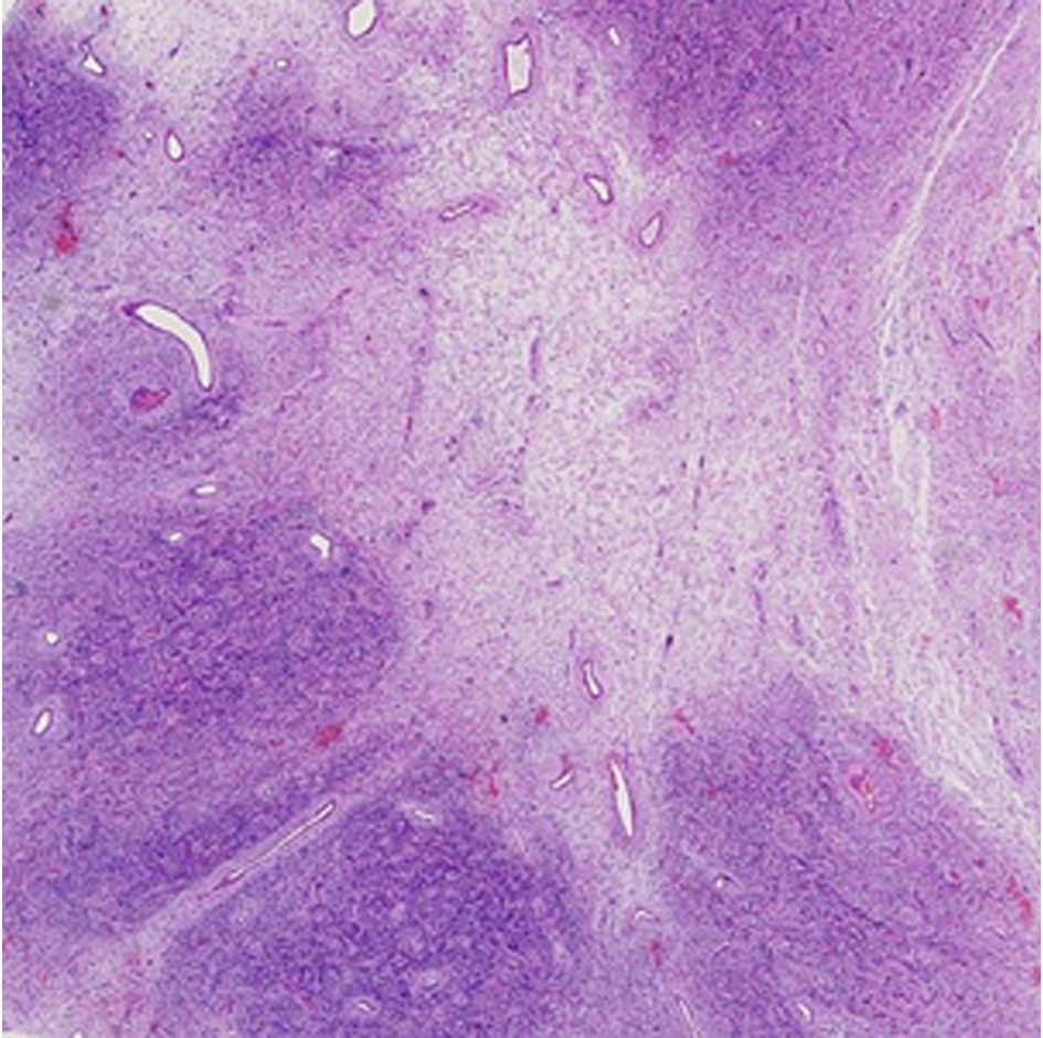
Figure 3 Histopathological examination by hematoxylin-eosin staining (20 ×).
The tumor cells grew as mixed nodules in dense areas and sparse areas.
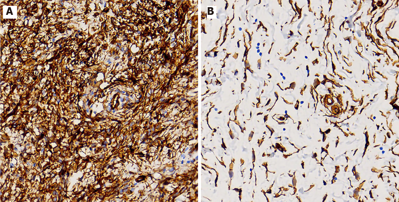
Figure 4 Immunohistochemical examination by the EnVision method (400 ×).
A: Diffuse and strong expression of CD34 in the dense area; B: The expression of CD34 was positive in the sparse area.
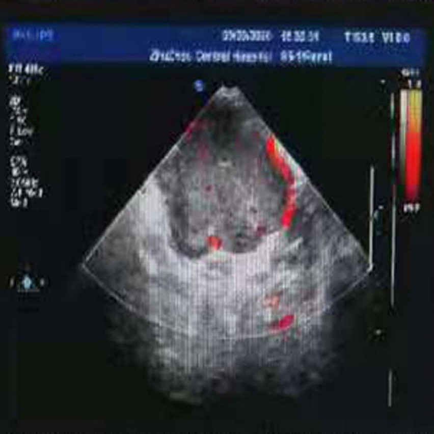
Figure 5 Ultrasound image.
A perianal cystic mass, which was initially considered as a perianal abscess, was observed.
- Citation: Long CY, Wang TL. Perianal superficial CD34-positive fibroblastic tumor: A case report. World J Clin Cases 2021; 9(20): 5605-5610
- URL: https://www.wjgnet.com/2307-8960/full/v9/i20/5605.htm
- DOI: https://dx.doi.org/10.12998/wjcc.v9.i20.5605


