Copyright
©The Author(s) 2021.
World J Clin Cases. Jul 16, 2021; 9(20): 5562-5567
Published online Jul 16, 2021. doi: 10.12998/wjcc.v9.i20.5562
Published online Jul 16, 2021. doi: 10.12998/wjcc.v9.i20.5562
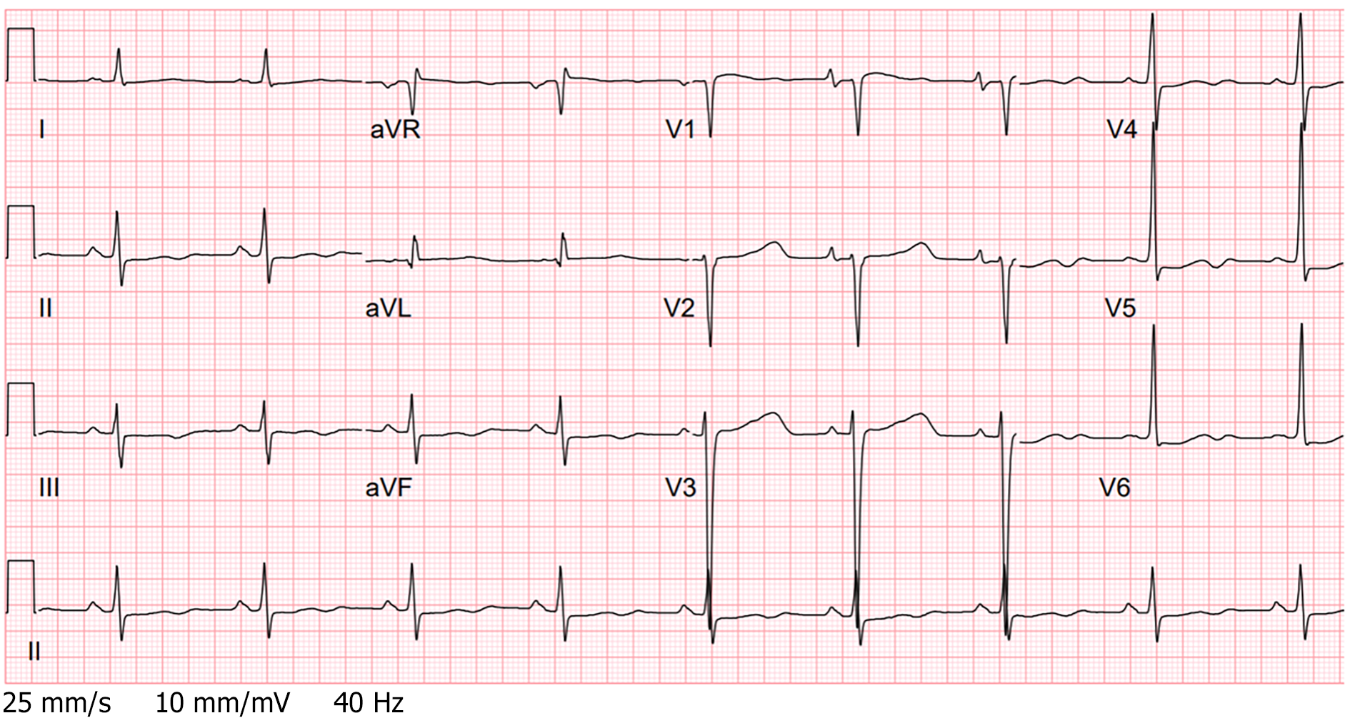
Figure 1 Electrocardiogram (prior to pacing wire placement): Sinus bradycardia and prolonged QTc interval of 516 milliseconds.
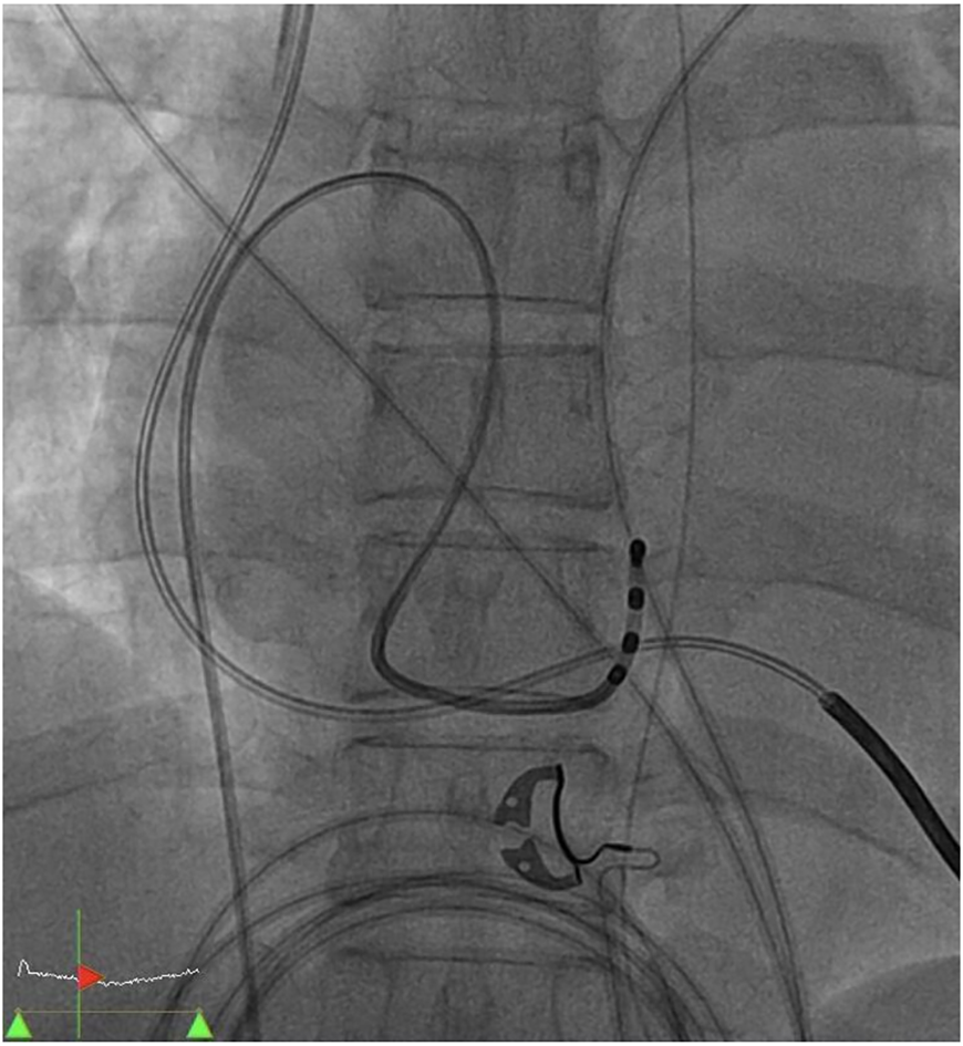
Figure 2 Cardiac catheterization: Placement of temporary pacing wire to the coronary sinus.
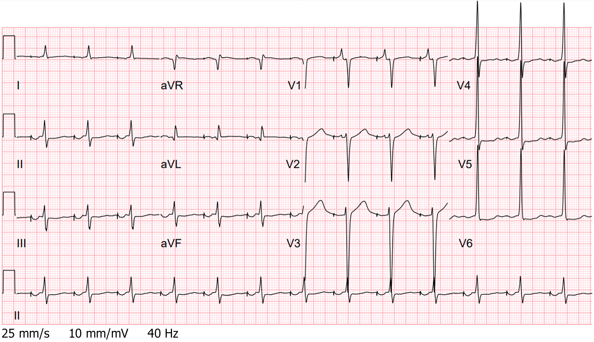
Figure 3 Electrocardiogram (after pacing wire placement): Atrial pacing with an AOO mode.
- Citation: Ju TR, Tseng H, Lin HT, Wang AL, Lee CC, Lai YC. Temporary coronary sinus pacing to improve ventricular dyssynchrony with cardiogenic shock: A case report. World J Clin Cases 2021; 9(20): 5562-5567
- URL: https://www.wjgnet.com/2307-8960/full/v9/i20/5562.htm
- DOI: https://dx.doi.org/10.12998/wjcc.v9.i20.5562


