Copyright
©The Author(s) 2021.
World J Clin Cases. Jun 26, 2021; 9(18): 4741-4747
Published online Jun 26, 2021. doi: 10.12998/wjcc.v9.i18.4741
Published online Jun 26, 2021. doi: 10.12998/wjcc.v9.i18.4741
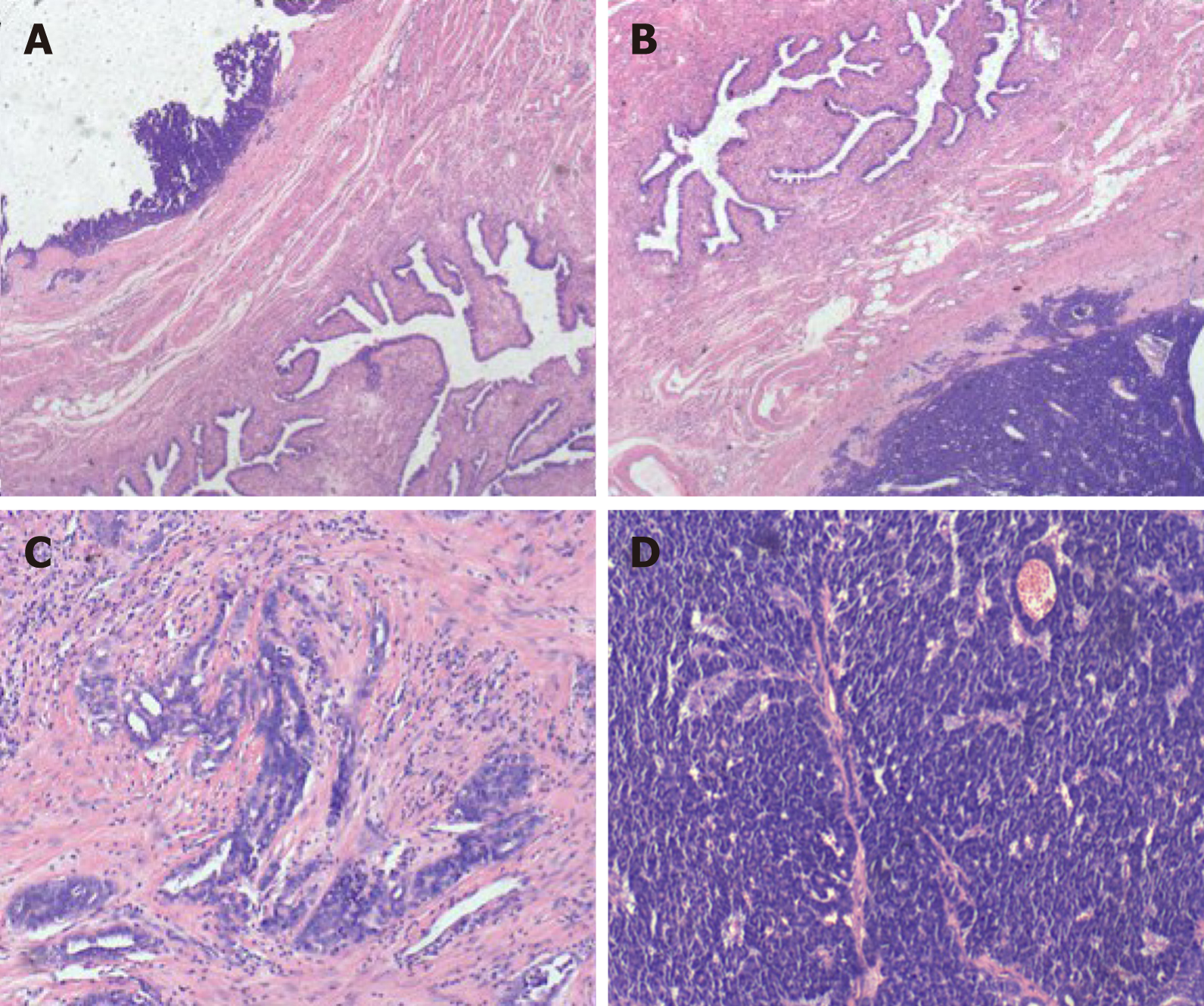
Figure 1 Histologic features of fallopian tube-mesonephric adenocarcinoma.
A and B: Pathological finding revealed that the tumor originated from the fallopian tube wall and involved the mucosa and serosal membrane of the fallopian tube; C and D: Vestiges of mesonephric hyperplasia (C) and hyperplasia into cancerous nests (D) were histologically found. The histologic patterns of the tumor were mainly solid.
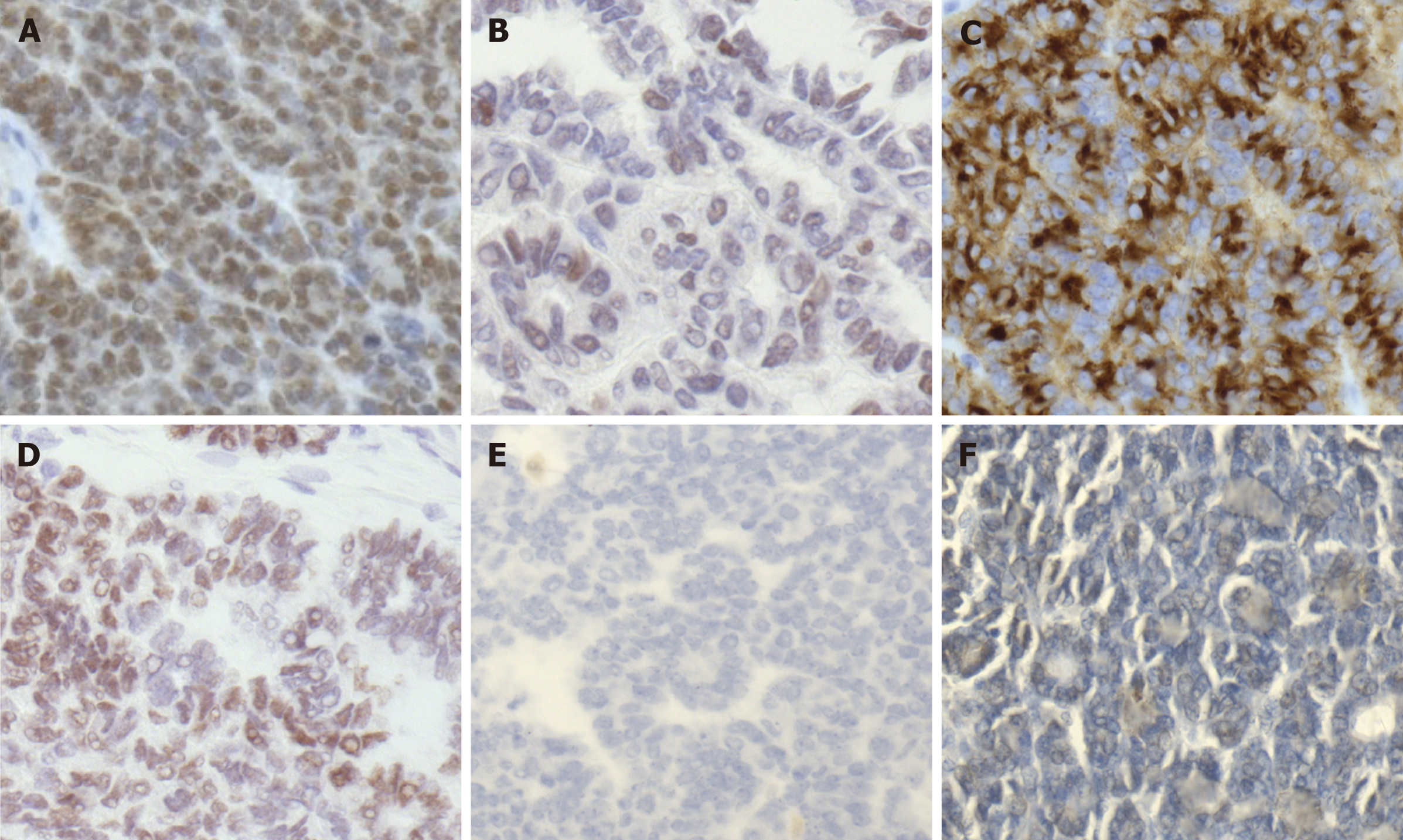
Figure 2 Immunohistochemical staining of fallopian tube-mesonephric adenocarcinoma.
A: PAX8 expression in this case was strong and diffuse, indicating that the tumor arose from the female genital tract; B: GATA3 positivity is helpful in confirming the diagnosis of mesonephric carcinoma, but the staining may be weak and focal. In the present case, the staining for GATA3 was weak and diffuse; C: CD10 revealed cytoplasmic and luminal staining; D: Immunohistochemical staining for thyroid transcription factor-1 was strong and diffuse; E and F: Immunohistochemistry showed totally negative staining for Calretinin (E) and Wilms' tumour-1 (F).
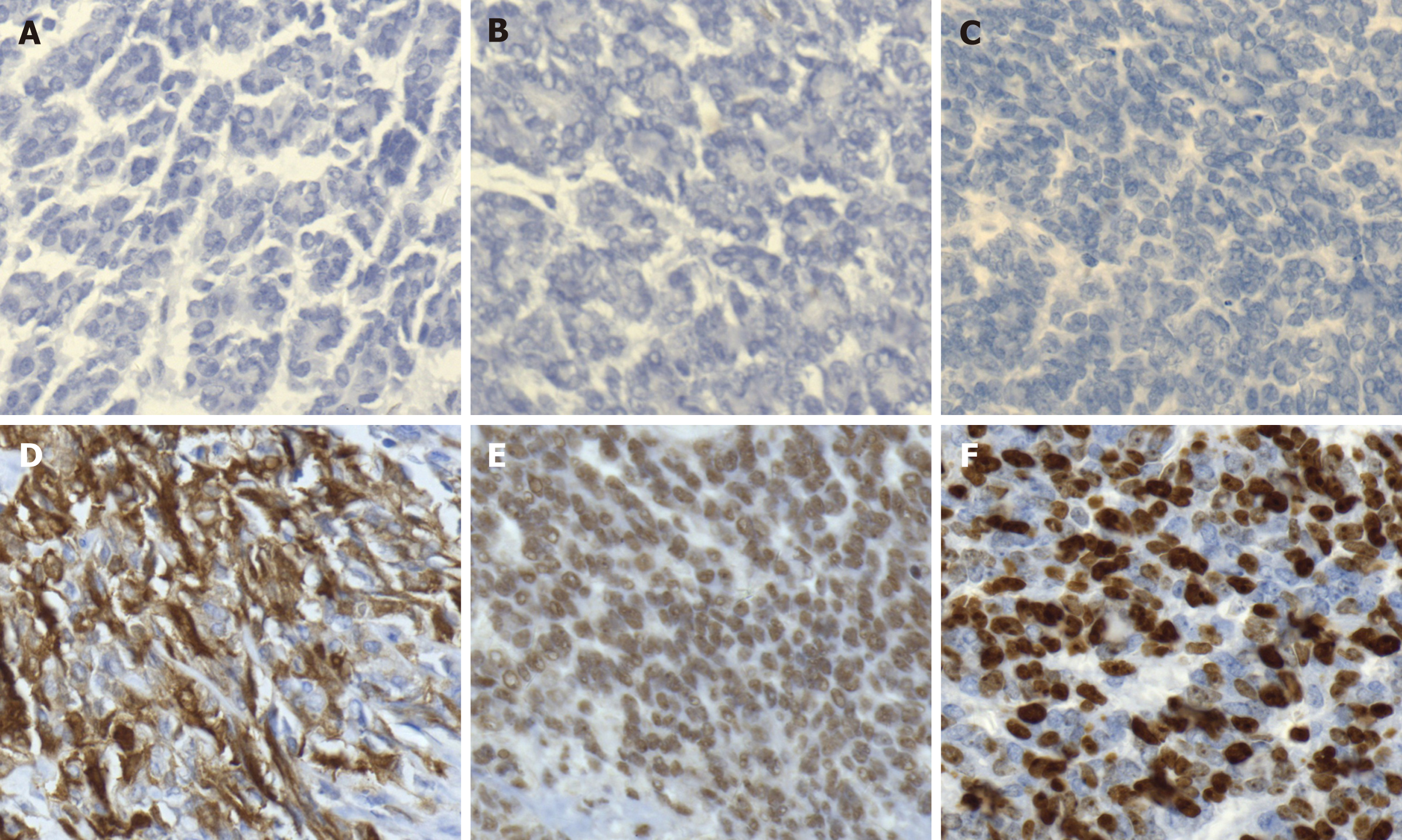
Figure 3 Immunohistochemical staining of fallopian tube-mesonephric adenocarcinoma.
A-C: Immunohistochemistry demonstrated totally negative staining for estrogen receptor (A), progesterone receptor (B), and CA125 (C); D and E: Positive staining for P16 (D) and P53 (E) was detected; F: The positive rate of Ki-67 expression was 60%-70%.
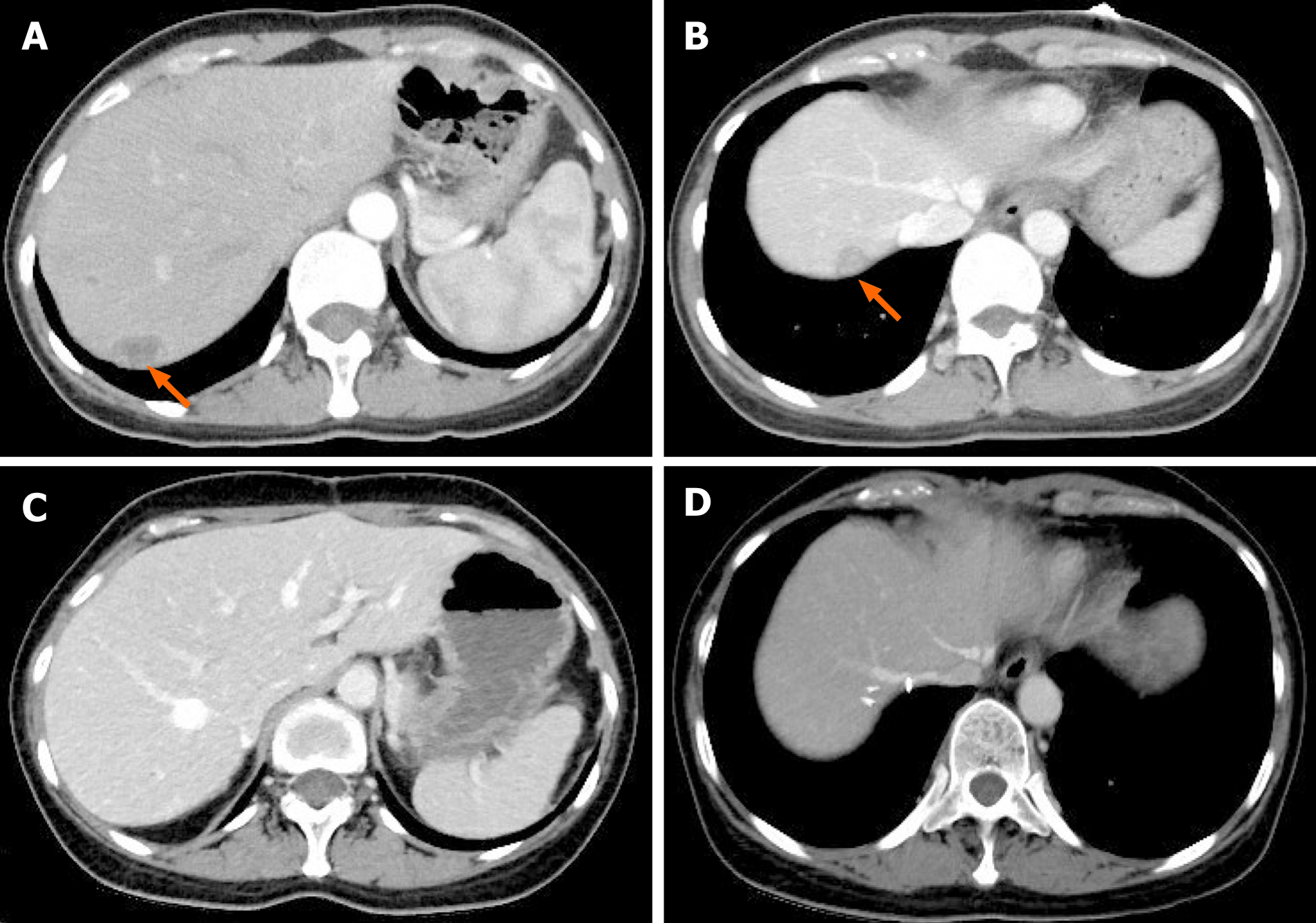
Figure 4 Computed tomography images of the patient with hepatic recurrence.
A and B: Abdominal computed tomography (CT) scan detected low-density nodules under the liver capsule. The arrows indicate metastatic nodules in the liver, and the diameters of the metastatic lesions ranged from 1.5-2.5 cm; C and D: CT appearances of the liver after second resection of tumors in the liver plus partial hepatectomy.
- Citation: Xie C, Shen YM, Chen QH, Bian C. Primary mesonephric adenocarcinoma of the fallopian tube: A case report. World J Clin Cases 2021; 9(18): 4741-4747
- URL: https://www.wjgnet.com/2307-8960/full/v9/i18/4741.htm
- DOI: https://dx.doi.org/10.12998/wjcc.v9.i18.4741


