Copyright
©The Author(s) 2021.
World J Clin Cases. Jun 6, 2021; 9(16): 3838-3847
Published online Jun 6, 2021. doi: 10.12998/wjcc.v9.i16.3838
Published online Jun 6, 2021. doi: 10.12998/wjcc.v9.i16.3838
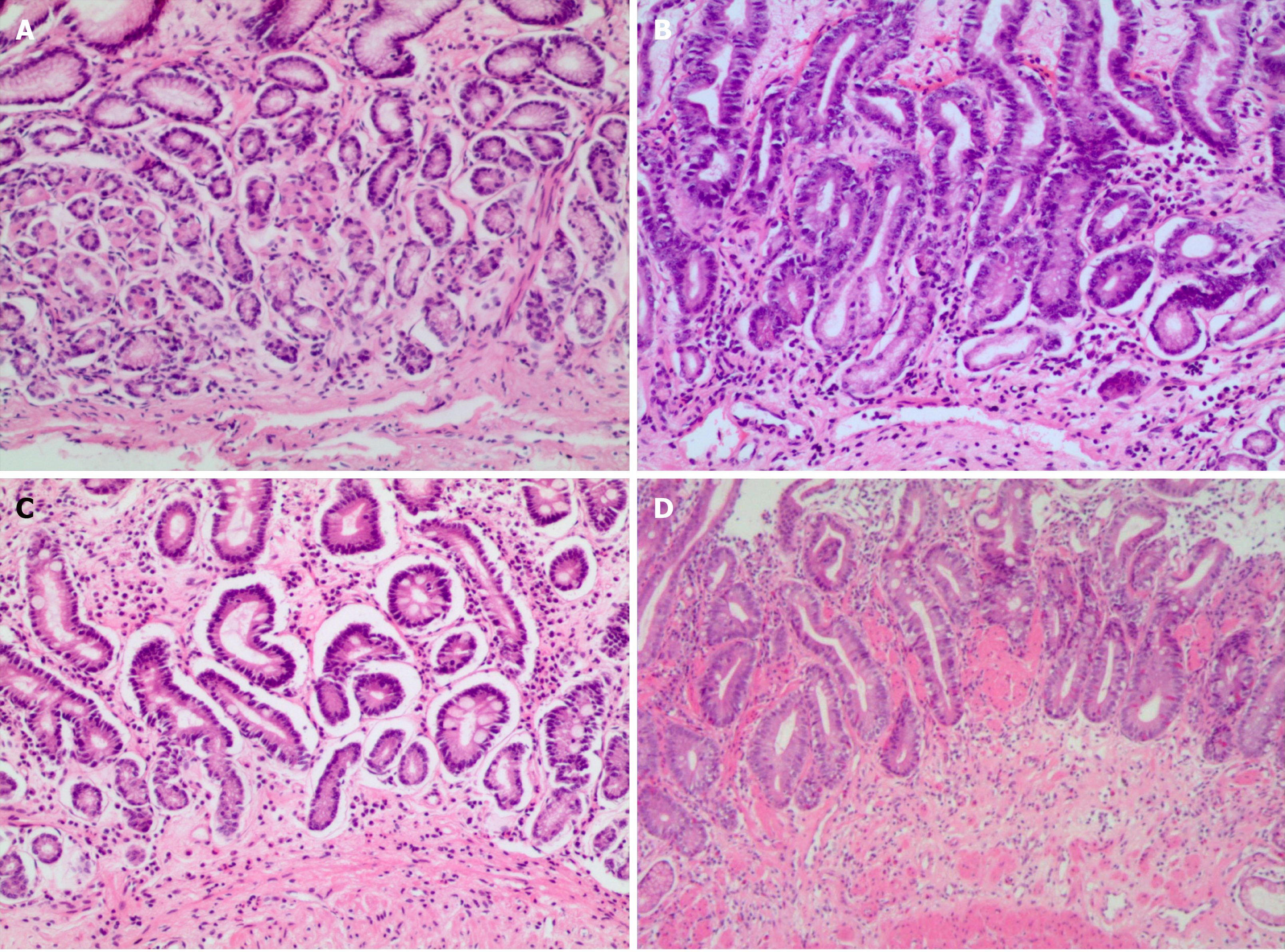
Figure 1 Histological analysis.
A: Simple type chronic atrophic gastritis—only thinning mucosal and gland atrophy, no glandular hyperplasia or heterotypic and intestinal metaplasia. Magnification, 20 ×; B: Hyperplasia type chronic atrophic gastritis—inherent layer atrophy accompanied by glandular hyperplasia, mainly quantitative, with no epithelial atypical hyperplasia. Magnification, 20 ×; C: Intestinal metaplasia type chronic atrophic gastritis—intrinsic glandular atrophy accompanied by intestinal metaplasia, mainly small intestinal epithelial metaplasia. Magnification, 20 ×; D: Intraepithelial neoplasia type chronic atrophic gastritis—inherent layer atrophy with high grade intraepithelial neoplasia. Magnification, 20 ×.
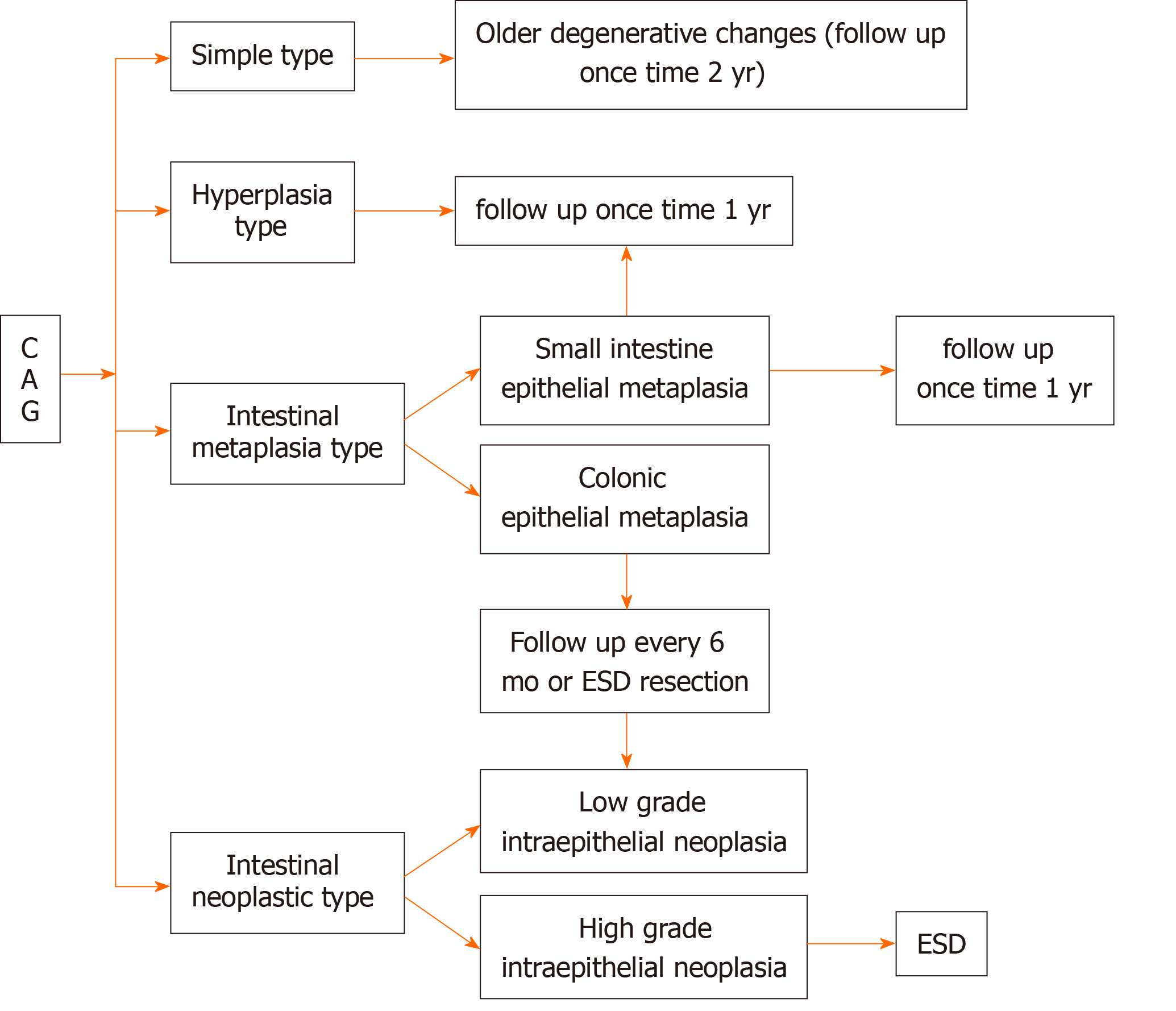
Figure 2 Histopathological typing and follow-up time of chronic atrophic gastritis.
CAG: Chronic atrophic gastritis; ESD: Endoscopic submucosal dissection.
- Citation: Wang YK, Shen L, Yun T, Yang BF, Zhu CY, Wang SN. Histopathological classification and follow-up analysis of chronic atrophic gastritis. World J Clin Cases 2021; 9(16): 3838-3847
- URL: https://www.wjgnet.com/2307-8960/full/v9/i16/3838.htm
- DOI: https://dx.doi.org/10.12998/wjcc.v9.i16.3838


