Copyright
©The Author(s) 2021.
World J Clin Cases. May 26, 2021; 9(15): 3765-3772
Published online May 26, 2021. doi: 10.12998/wjcc.v9.i15.3765
Published online May 26, 2021. doi: 10.12998/wjcc.v9.i15.3765
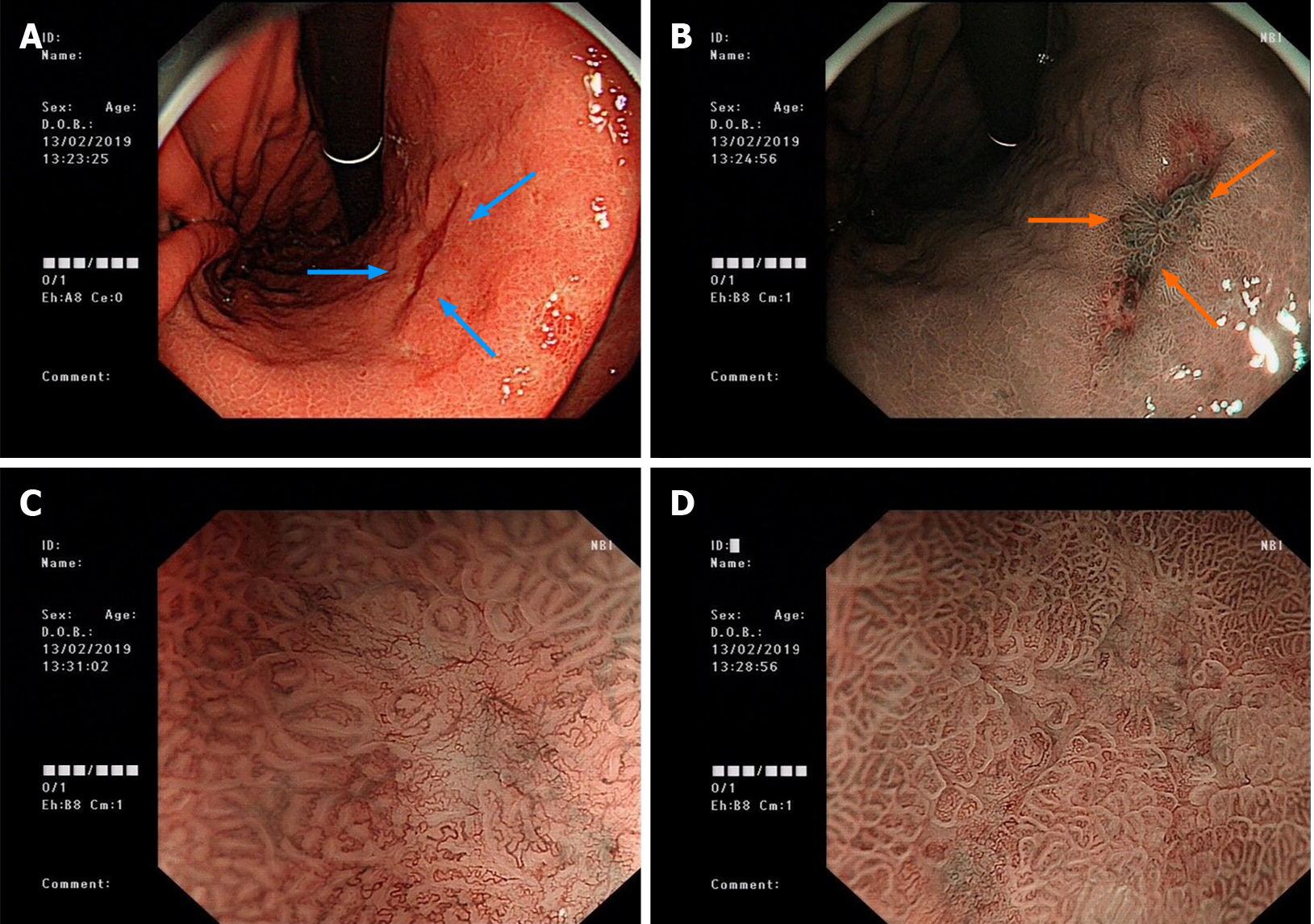
Figure 1 Images of electronic gastroscopy before the gastrectomy.
A: Image showing a 2.0 cm × 1.0 cm flat concave mucosa on the lesser curvature of the lower stomach (blue arrows); B: Image of narrow band imaging; C and D: Images of magnifying gastroscopy.
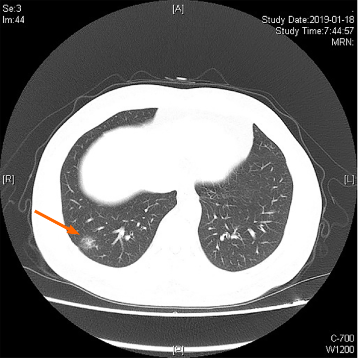
Figure 2 Images of chest computed tomography before the resection of the right lower lobe of the right lung, which showed a nodule in the lower lobe of the right lung (orange arrow).
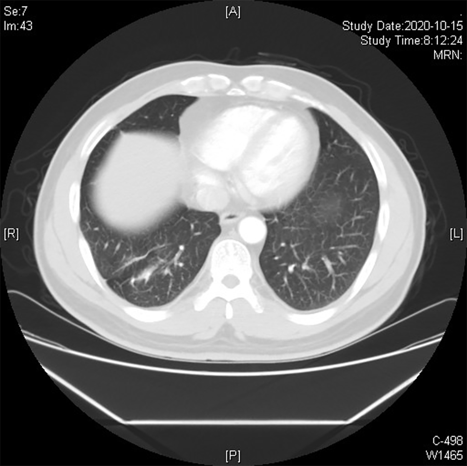
Figure 3 No metastasis or recurrence was found on follow-up chest computed tomography, 16 mo after the resection operation.
Figure 4 No metastasis or recurrence was found in the gastroscopy of the upper abdomen, 10 mo after radical distal gastrectomy.
- Citation: Rao W, Liu FG, Jiang YP, Xie M. De novo multiple primary carcinomas in a patient after liver transplantation: A case report. World J Clin Cases 2021; 9(15): 3765-3772
- URL: https://www.wjgnet.com/2307-8960/full/v9/i15/3765.htm
- DOI: https://dx.doi.org/10.12998/wjcc.v9.i15.3765


