Copyright
©The Author(s) 2021.
World J Clin Cases. May 16, 2021; 9(14): 3478-3486
Published online May 16, 2021. doi: 10.12998/wjcc.v9.i14.3478
Published online May 16, 2021. doi: 10.12998/wjcc.v9.i14.3478
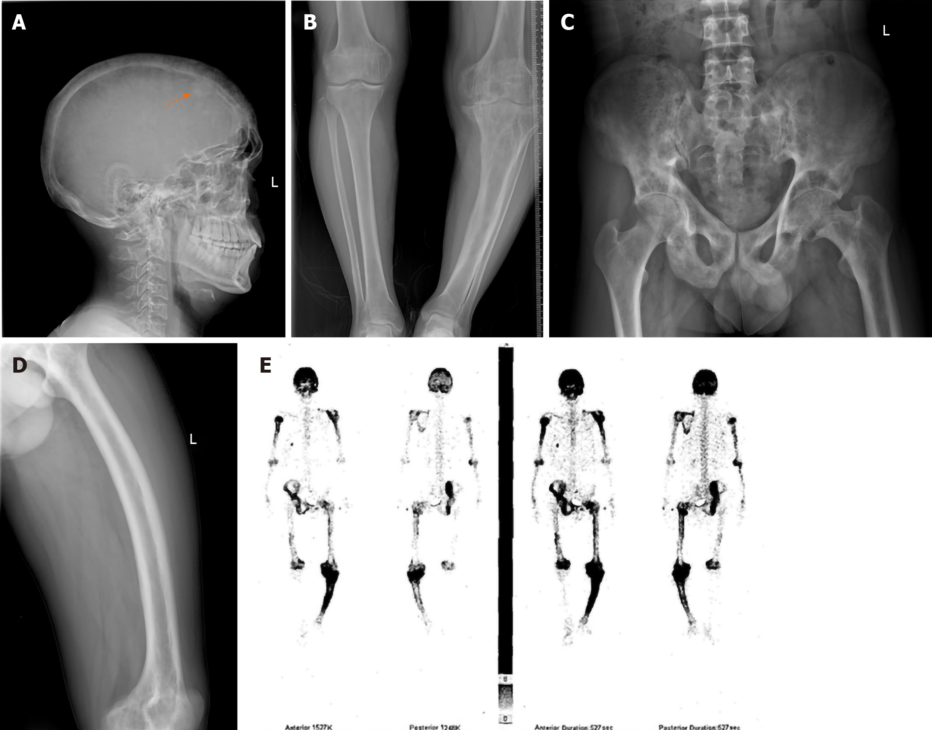
Figure 1 Imaging changes in patients with Paget’s disease of bone.
A: Radiograph of case 8 showing bone lesions in the skull and cranial plate and a slightly thickened, localized high-density “cotton ball” appearance (arrowhead); B: X-ray of the lower limbs from case 8 showing diffuse bone lesions in the left femur, tibia, and fibula, a thickened bone cortex, uneven density in the marrow cavity, and expanded femur and tibia metaphyses; C: X-ray of the hip from case 10 showing multiple cystic radiolucent spaces in the pelvic bones; D: Radiograph of the left femur from case 10 showing a thickened bone cortex, narrowed marrow cavity, and sabre-like deformation of the femur; E: Technetium-99 conjugated with methylene diphosphonate bone scans from case 8 showing increased uptake of radionuclide in the skull, left scapula, right fifth anterior rib, and right hemipelvis and limb bones.
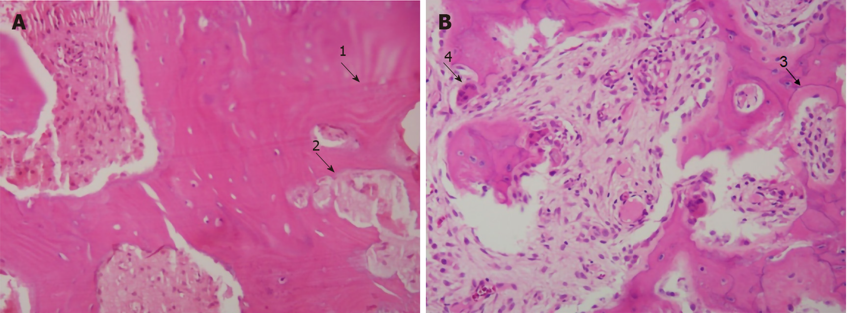
Figure 2 Histopathological features of Paget’s disease of bone.
A: Biopsy specimen from case 9 showing irregular broad bone trabeculae (arrowhead 1) and fibrous vascular tissue (arrowhead 2); B: Pathological tissue from case 11 showing a mosaic appearance with irregularly arranged cement lines (arrowhead 3) and multinuclear osteoclasts (arrowhead 4) (hematoxylin-eosin staining, magnification × 200).
- Citation: Miao XY, Wang XL, Lyu ZH, Ba JM, Pei Y, Dou JT, Gu WJ, Du J, Guo QH, Chen K, Mu YM. Paget’s disease of bone: Report of 11 cases. World J Clin Cases 2021; 9(14): 3478-3486
- URL: https://www.wjgnet.com/2307-8960/full/v9/i14/3478.htm
- DOI: https://dx.doi.org/10.12998/wjcc.v9.i14.3478


