Copyright
©The Author(s) 2021.
World J Clin Cases. May 16, 2021; 9(14): 3458-3465
Published online May 16, 2021. doi: 10.12998/wjcc.v9.i14.3458
Published online May 16, 2021. doi: 10.12998/wjcc.v9.i14.3458
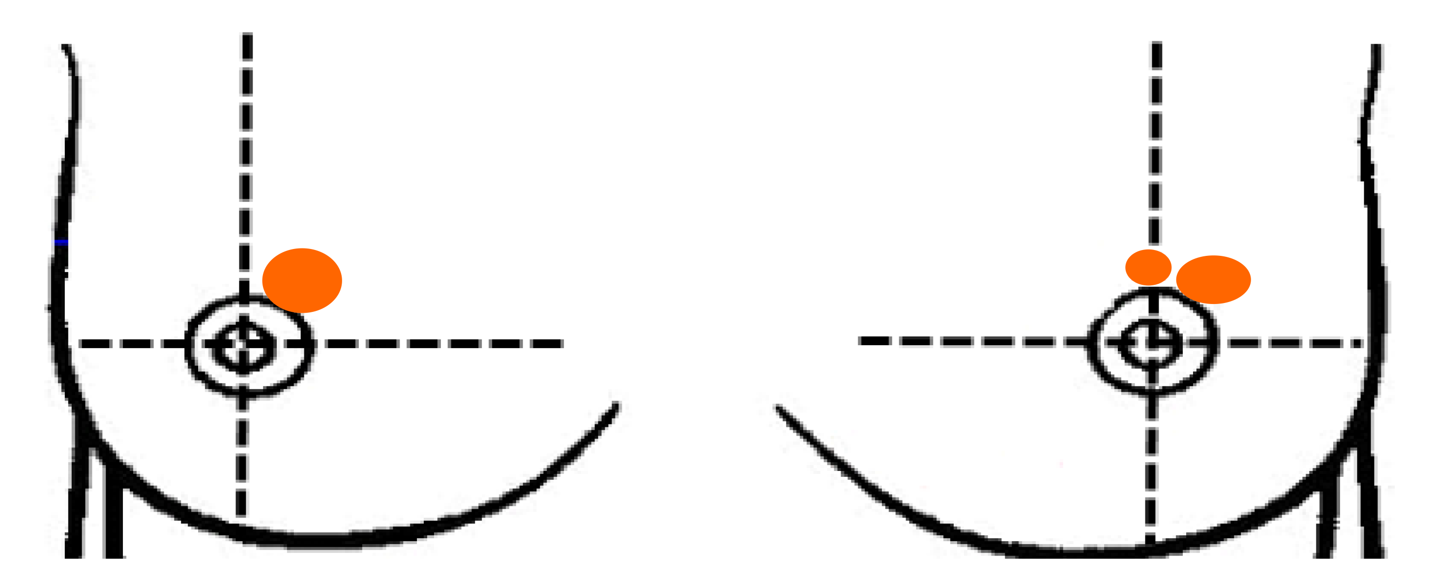
Figure 1 Schematic diagram of breast masses.
Three separate masses of the breast, including two solid masses in the left breast and one hard mass in the right breast, were palpable.
Figure 2 Ultrasonographic images of bilateral breasts.
There is a very hypoechoic zone at the 1 o'clock position of the right breast and the left breast, about 19.2 mm × 14.7 mm × 17.1 mm and 15.9 mm × 10.5 mm × 9.5 mm in size, respectively, irregular in shape, with an unclear border. There is a hypoechoic area at the 12 o'clock area in the left breast, about 5.9 mm × 6.0 mm × 5.9 mm, irregular in shape, with an unclear boundary.
Figure 3 Axial enhanced magnetic resonance images of bilateral breasts.
Cross-sectional contrast-enhanced FT1WI showed that focal and heterogeneous non-mass enhancement in the upper inner quadrant of the right breast and in the upper outer quadrant of the left breast.
Figure 4 Histopathological images (hematoxylin-eosin staining).
A and B: At high and medium magnification, dense lymphocytes infiltrating around the lobules of the breast, and extensive fibrosis of the surrounding stroma were observed; C: At medium magnification, myofibroblasts presented epithelioid changes.
- Citation: Chen XX, Shao SJ, Wan H. Diabetic mastopathy in an elderly woman misdiagnosed as breast cancer: A case report and review of the literature. World J Clin Cases 2021; 9(14): 3458-3465
- URL: https://www.wjgnet.com/2307-8960/full/v9/i14/3458.htm
- DOI: https://dx.doi.org/10.12998/wjcc.v9.i14.3458


