Copyright
©The Author(s) 2021.
World J Clin Cases. Apr 26, 2021; 9(12): 2890-2898
Published online Apr 26, 2021. doi: 10.12998/wjcc.v9.i12.2890
Published online Apr 26, 2021. doi: 10.12998/wjcc.v9.i12.2890
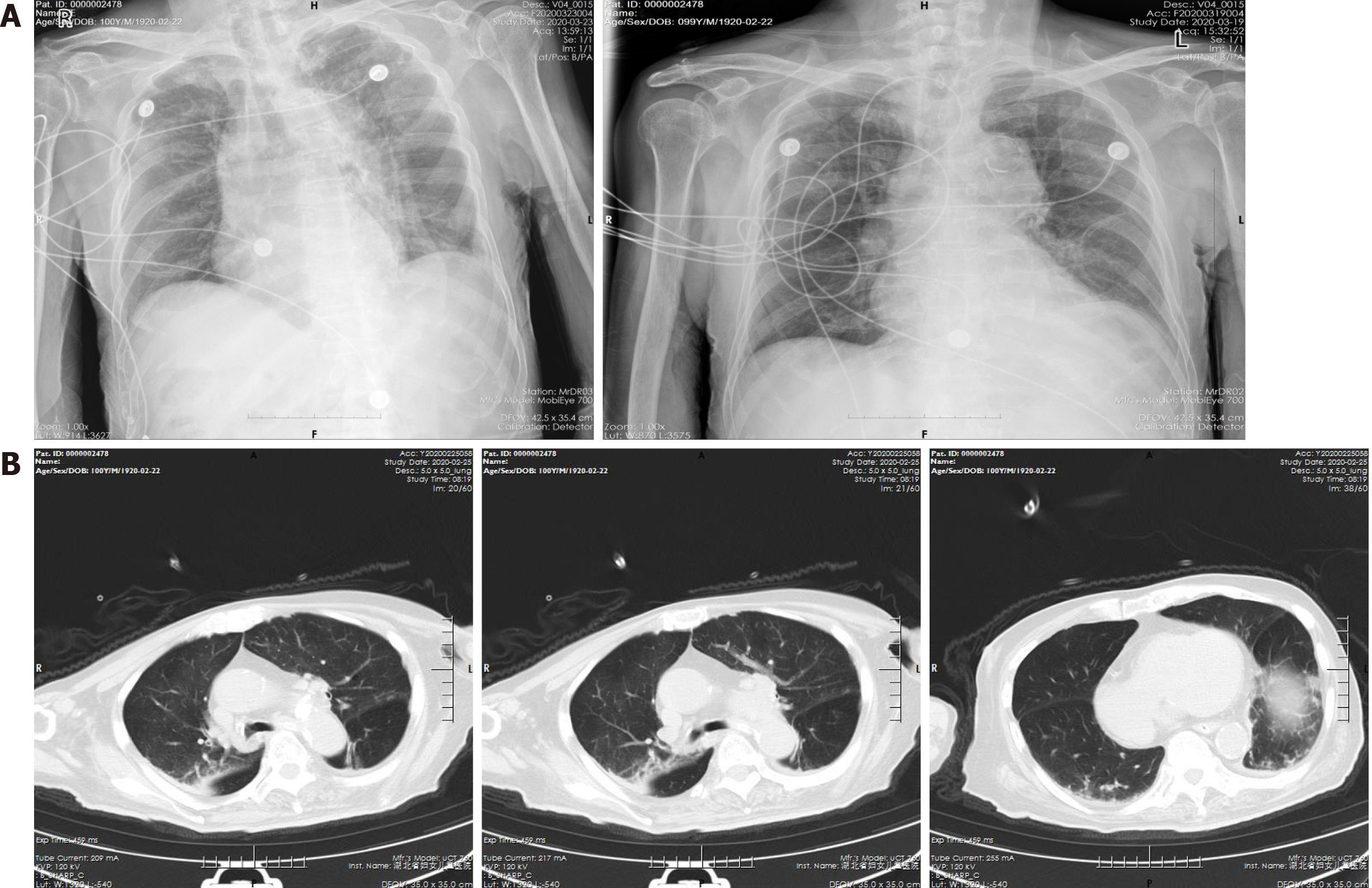
Figure 1 Anterior-posterior chest radiograph and computed tomography chest scan on February 25, 2020 (day 2 of hospitalization).
A: Anterior-posterior chest radiograph; B: Computed tomography chest scan. Small amounts of abnormally dense shadows in both lungs, suggesting infectious pneumonia; chronic bronchitis with pulmonary emphysema; old left upper lung lesion; bilateral pleural hypertrophy and calcification; aortic atherosclerosis; and small cyst of the left liver.
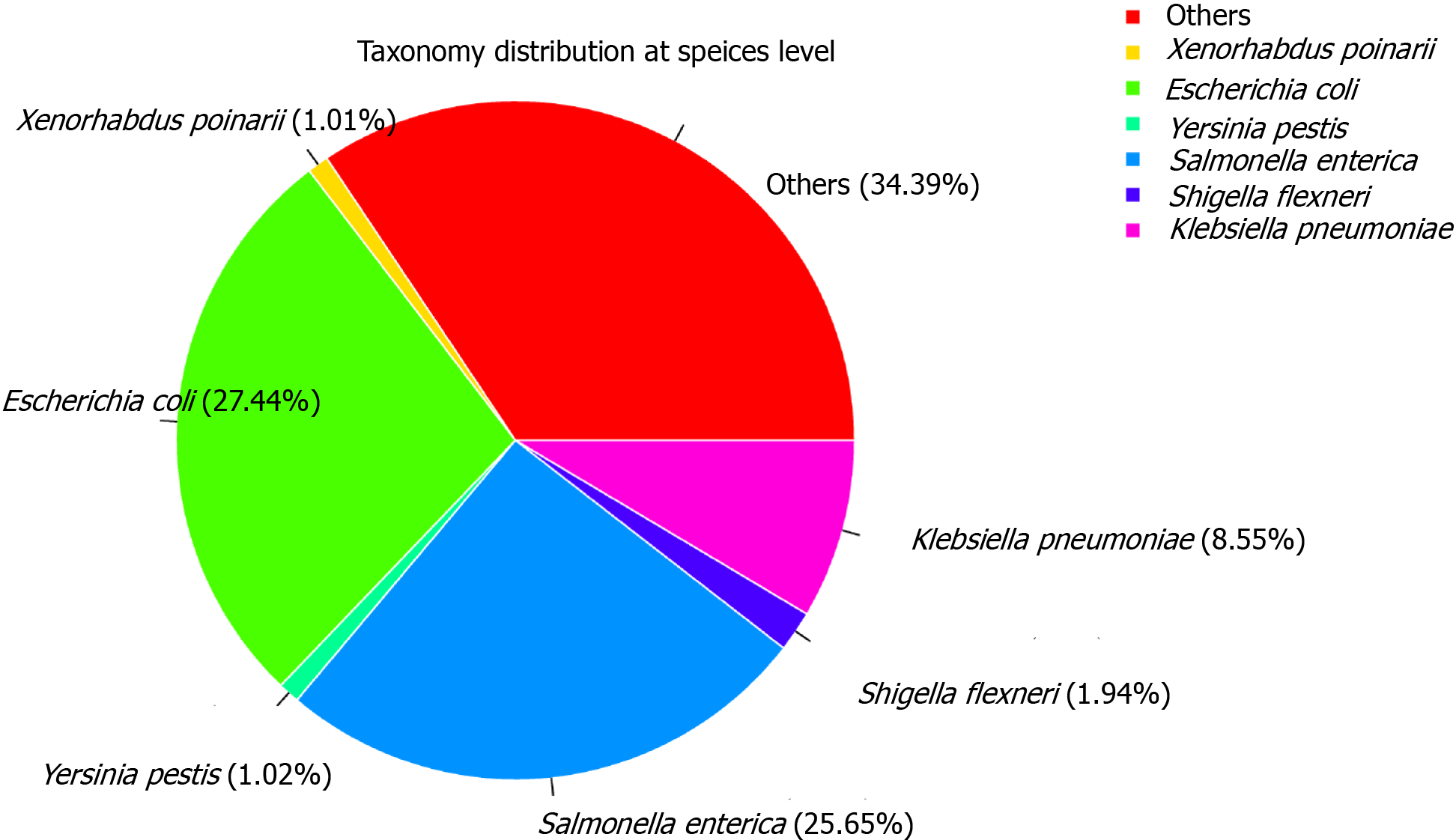
Figure 2 Visualization of the taxonomy classification interactively.
Filtered sequences from the anal swab sample were classified by Kraken 2 software and visualized by using Krona tools (https://github.com/marbl/Krona). The distribution of each organism at each taxonomy level can be see interactively. At the domain level, 99.24% of the sequence were classified as bacteria and only 0.05% were classified as viruses.
- Citation: Liu B, Ren KK, Wang N, Xu XP, Wu J. Timing of convalescent plasma therapy-tips from curing a 100-year-old COVID-19 patient using convalescent plasma treatment: A case report . World J Clin Cases 2021; 9(12): 2890-2898
- URL: https://www.wjgnet.com/2307-8960/full/v9/i12/2890.htm
- DOI: https://dx.doi.org/10.12998/wjcc.v9.i12.2890


