Copyright
©The Author(s) 2020.
World J Clin Cases. Apr 26, 2020; 8(8): 1538-1546
Published online Apr 26, 2020. doi: 10.12998/wjcc.v8.i8.1538
Published online Apr 26, 2020. doi: 10.12998/wjcc.v8.i8.1538
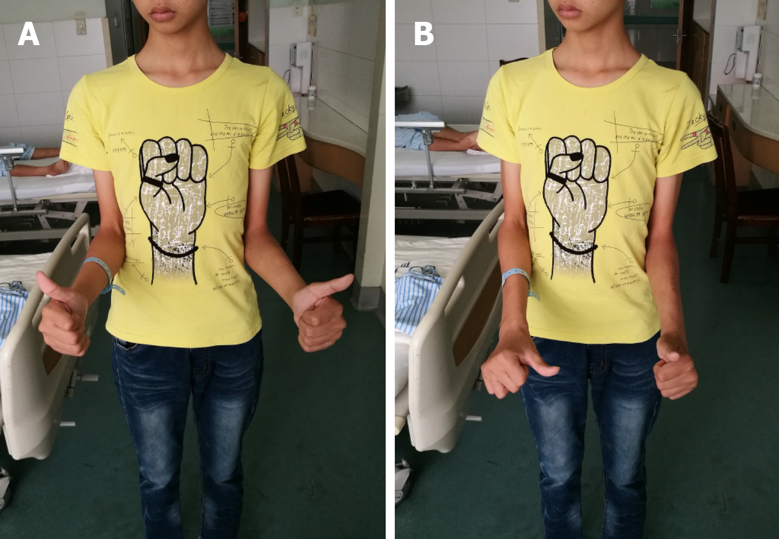
Figure 1 Active motion of the left forearm was limited.
In order to reduce the rotation error caused by the compensation of elbow motion, the elbow joint was tightly attached to the waist. A: Supination; B: Pronation.
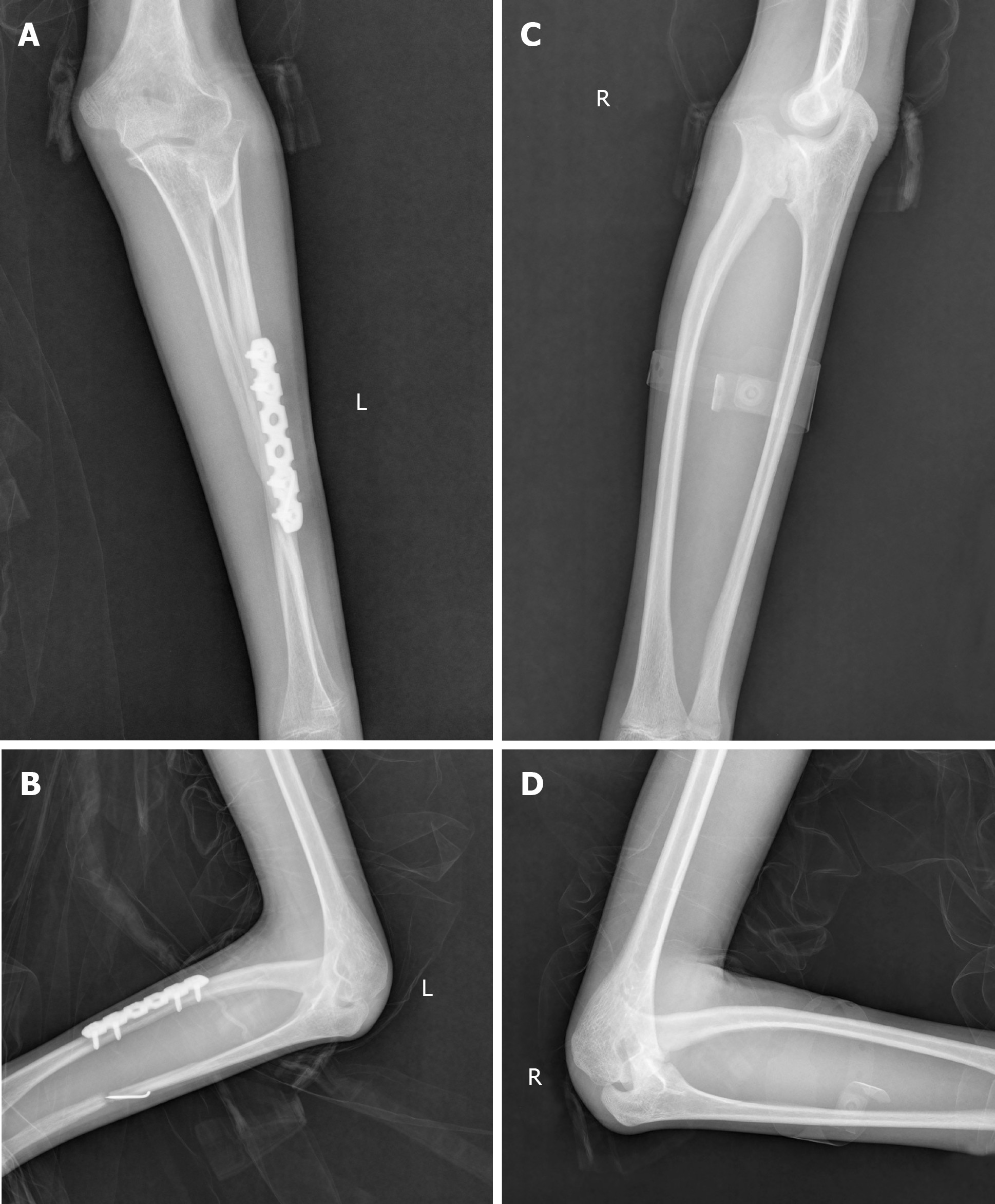
Figure 2 Plain radiographs of the bilateral forearm at 3 mo after surgery.
A, B: Left side with loss of reduced location; C, D: Right side with the same deformity of radioulnar synostosis.
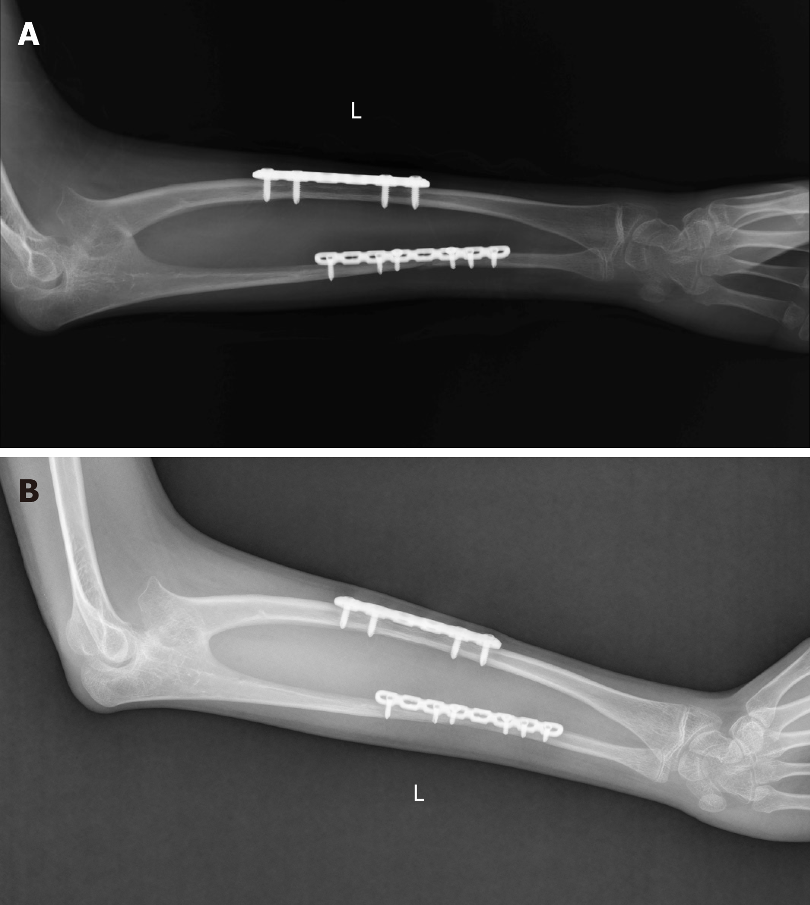
Figure 3 Plain radiographs showing the formation of callus, the disappearance of fracture line and the healing of fracture.
A: At 2 mo after surgery; B: At 1 yr after surgery.
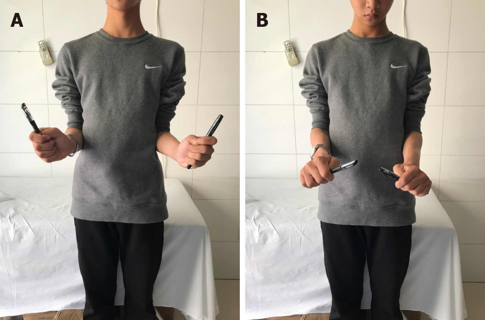
Figure 4 The range of motion of the left forearm was restored to the pre-injury state and was significantly improved after operation.
A: Supination; B: Pronation.
- Citation: Yang ZY, Ni JD, Long Z, Kuang LT, Tao SB. Unusual presentation of congenital radioulnar synostosis with osteoporosis, fragility fracture and nonunion: A case report and review of literature. World J Clin Cases 2020; 8(8): 1538-1546
- URL: https://www.wjgnet.com/2307-8960/full/v8/i8/1538.htm
- DOI: https://dx.doi.org/10.12998/wjcc.v8.i8.1538


