Copyright
©The Author(s) 2020.
World J Clin Cases. Dec 26, 2020; 8(24): 6243-6251
Published online Dec 26, 2020. doi: 10.12998/wjcc.v8.i24.6243
Published online Dec 26, 2020. doi: 10.12998/wjcc.v8.i24.6243
Figure 1 Mean increase in the best-corrected visual acuity from the pretreatment value.
BCVA: Best-corrected visual acuity.
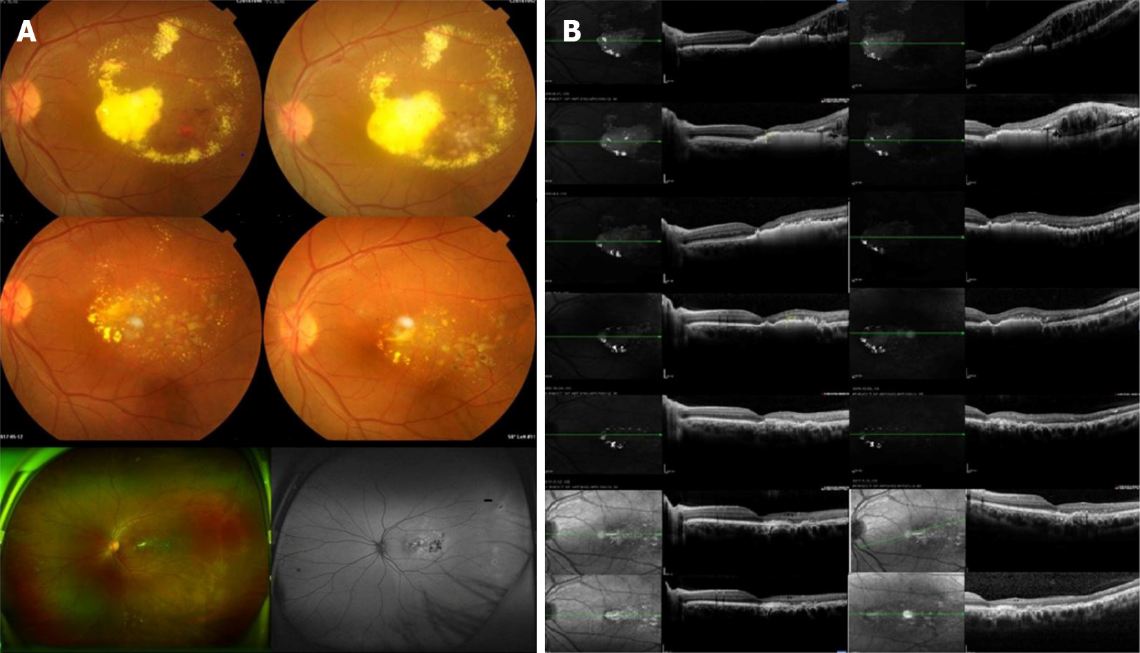
Figure 2 Coats disease of the left eye treated with conbercept combined with 532-laser photocoagulation.
A: Fundus photographs of the patient’s eye before treatment and at 1 mo, 1 yr and 2 yrs after treatment as well as at the last follow-up; B: Changes in central retinal thickness and subretinal fluid before treatment and at 1 wk, 1 mo, 3 mo, 1 yr and 2 yrs after treatment as well as at the final follow-up. The retinal interstitial layer and subretinal regions showed gradual reductions in edema and exudation.
- Citation: Jiang L, Qin B, Luo XL, Cao H, Deng TM, Yang MM, Meng T, Yang HQ. Three-year follow-up of Coats disease treated with conbercept and 532-nm laser photocoagulation. World J Clin Cases 2020; 8(24): 6243-6251
- URL: https://www.wjgnet.com/2307-8960/full/v8/i24/6243.htm
- DOI: https://dx.doi.org/10.12998/wjcc.v8.i24.6243


