Copyright
©The Author(s) 2020.
World J Clin Cases. Dec 6, 2020; 8(23): 6181-6189
Published online Dec 6, 2020. doi: 10.12998/wjcc.v8.i23.6181
Published online Dec 6, 2020. doi: 10.12998/wjcc.v8.i23.6181
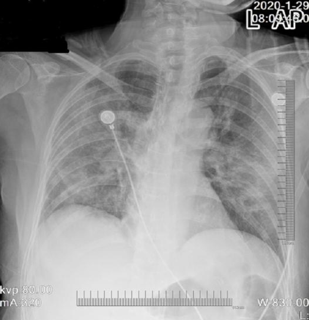
Figure 1 Chest radiograph revealing bilateral multipatchy consolidation and ground-glass opacities.
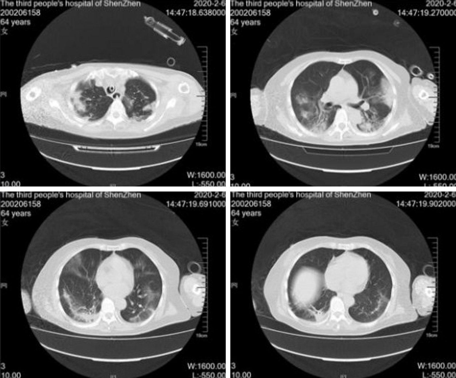
Figure 2 Chest computed tomography images revealing that exudate increased in the lung 11 d after admission.
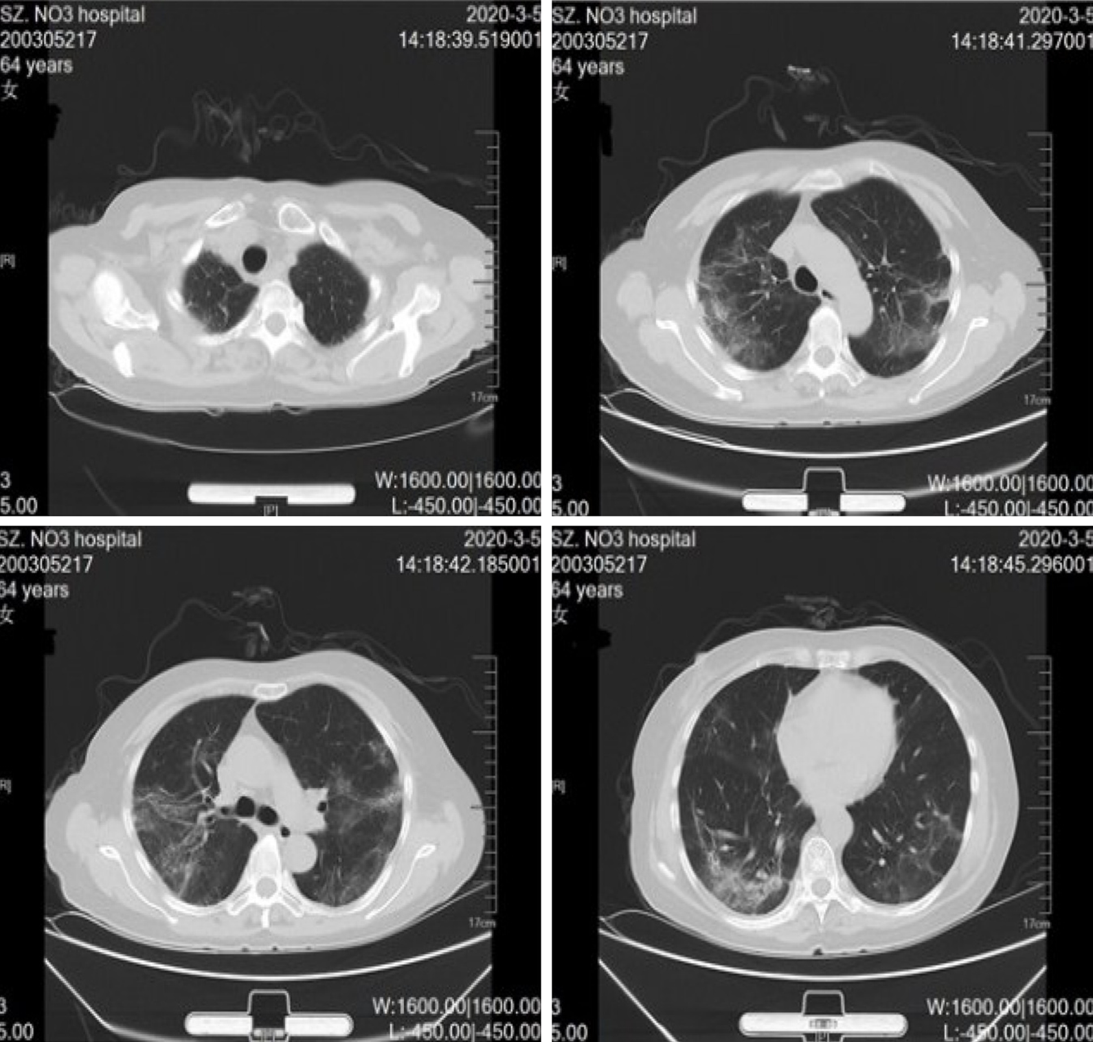
Figure 3 Chest computed tomography images showing almost complete resolution of infiltrates 39 d after admission.
- Citation: Pang QL, He WC, Li JX, Huang L. Symptomatic and optimal supportive care of critical COVID-19: A case report and literature review. World J Clin Cases 2020; 8(23): 6181-6189
- URL: https://www.wjgnet.com/2307-8960/full/v8/i23/6181.htm
- DOI: https://dx.doi.org/10.12998/wjcc.v8.i23.6181


