Copyright
©The Author(s) 2020.
World J Clin Cases. Dec 6, 2020; 8(23): 6158-6163
Published online Dec 6, 2020. doi: 10.12998/wjcc.v8.i23.6158
Published online Dec 6, 2020. doi: 10.12998/wjcc.v8.i23.6158
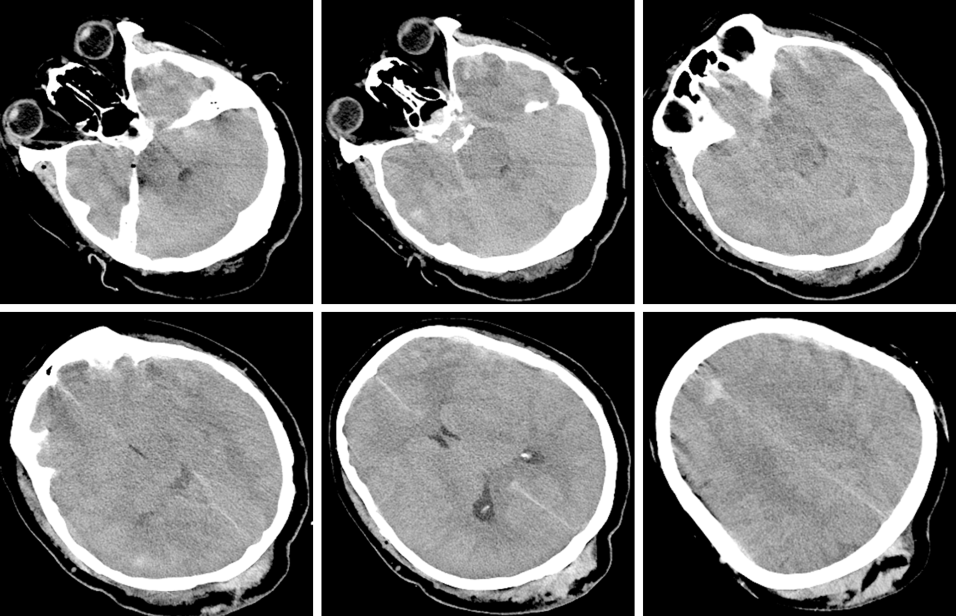
Figure 1 pecks of blood in the left frontal and temporal lobes.
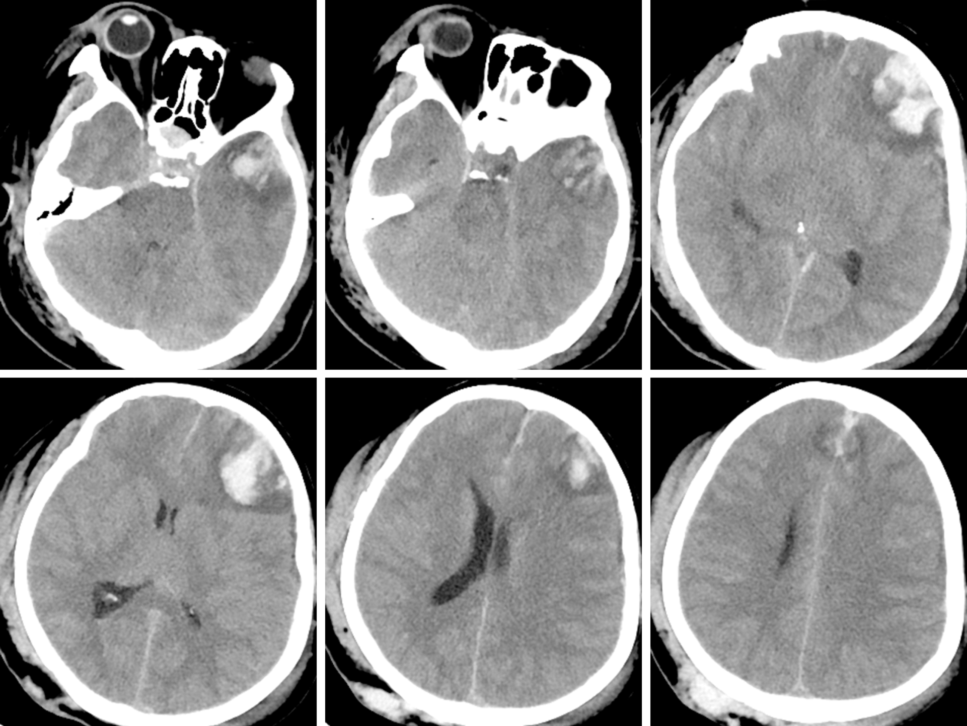
Figure 2 Haematomas in the left frontal and temporal lobes and 9 mm midline shift.
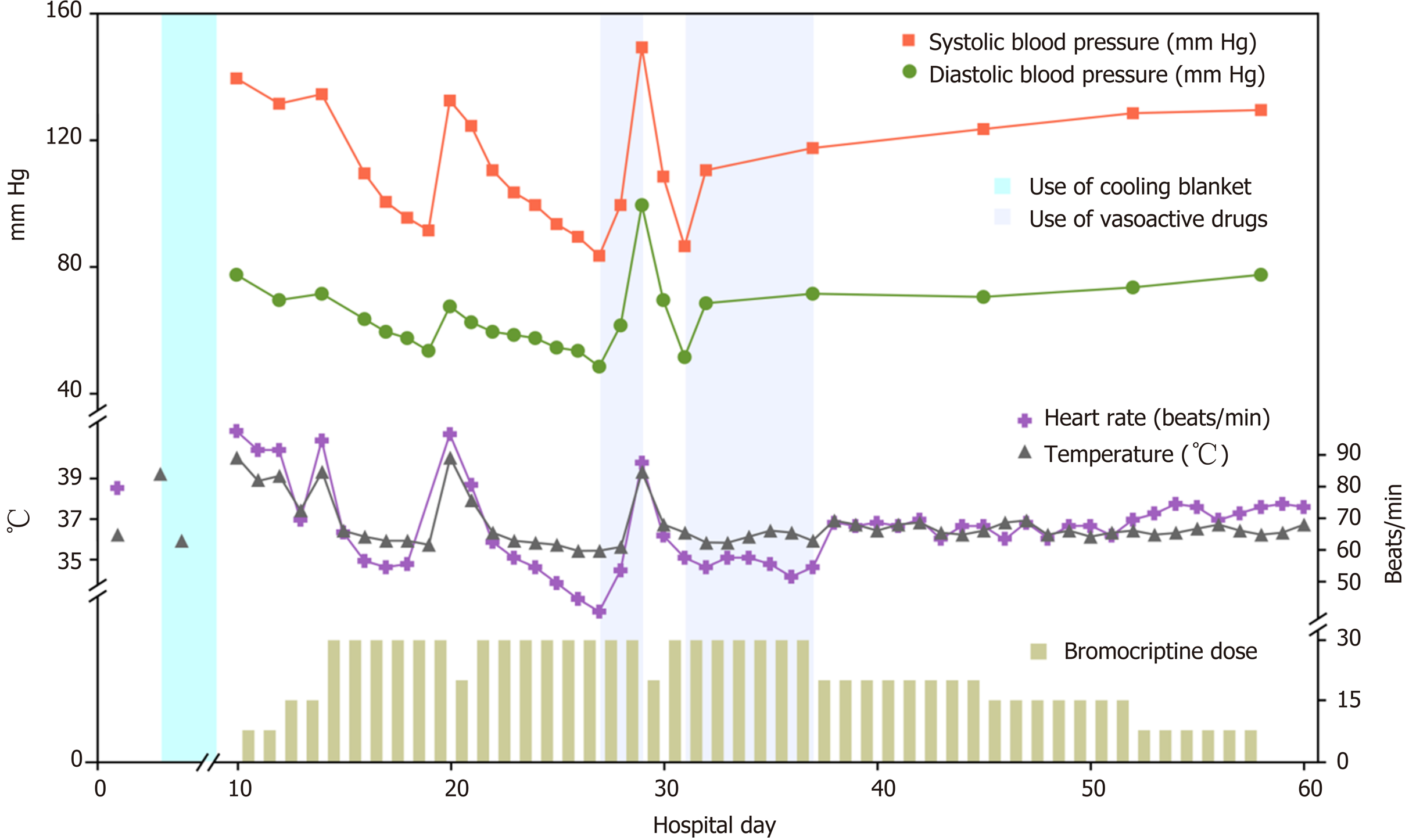
Figure 3 Fluctuations of marked temperature, blood pressure, heart rate, and dose of bromocriptine and treatments with cooling blanket and vasoactive drugs were noted during the clinical course.
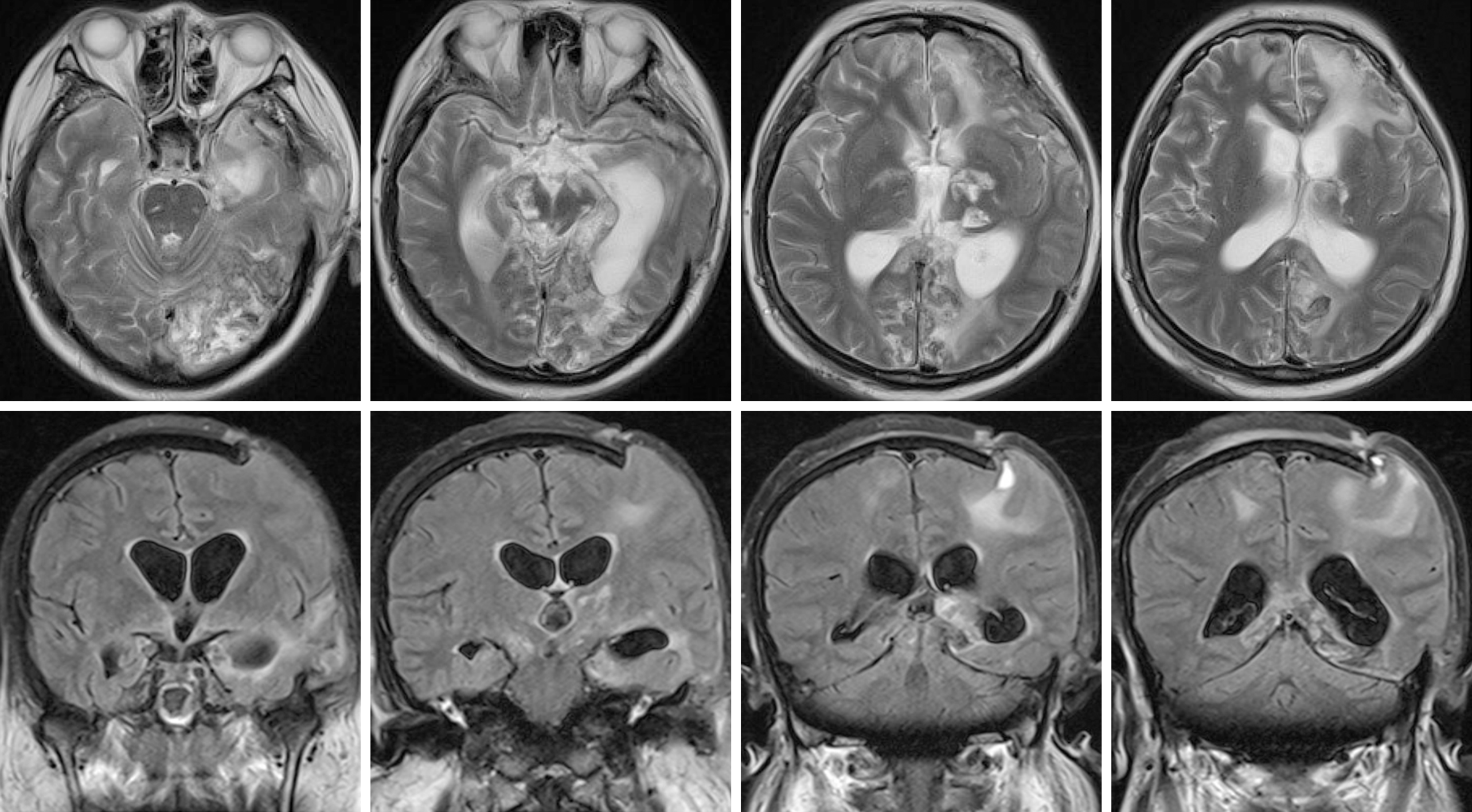
Figure 4 Speck signals in the midbrain, pontine, and pituitarium and multiple damage signals in the left frontal, temporal and occipital lobes, basal ganglia, thalamus, and corpus callosum.
- Citation: Ge X, Luan X. Uncontrolled central hyperthermia by standard dose of bromocriptine: A case report. World J Clin Cases 2020; 8(23): 6158-6163
- URL: https://www.wjgnet.com/2307-8960/full/v8/i23/6158.htm
- DOI: https://dx.doi.org/10.12998/wjcc.v8.i23.6158


