Copyright
©The Author(s) 2020.
World J Clin Cases. Dec 6, 2020; 8(23): 5944-5951
Published online Dec 6, 2020. doi: 10.12998/wjcc.v8.i23.5944
Published online Dec 6, 2020. doi: 10.12998/wjcc.v8.i23.5944
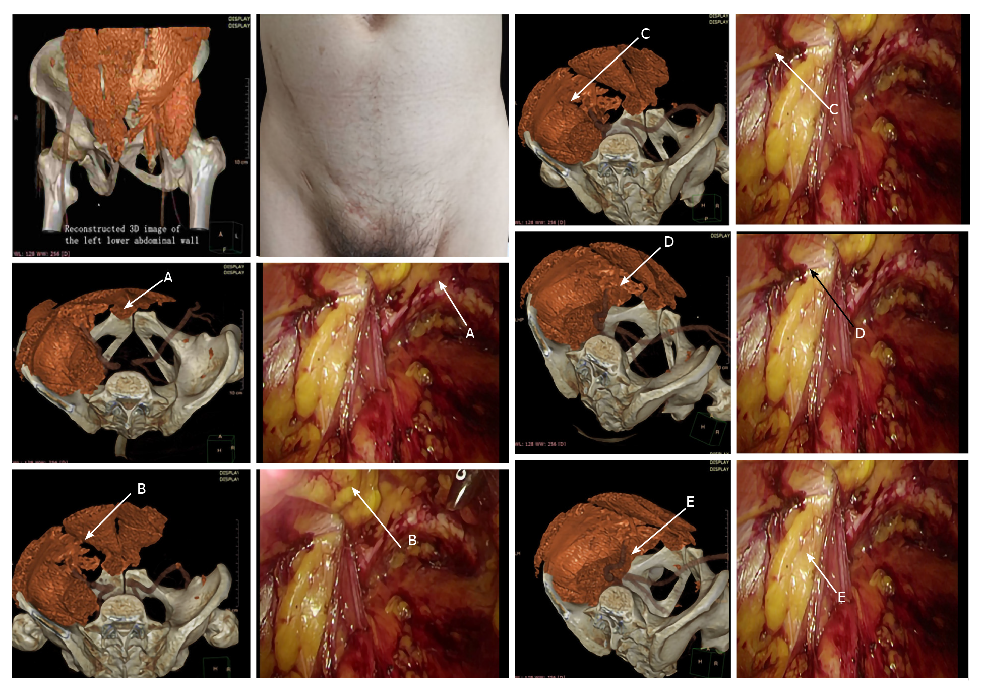
Figure 1 Comparison of the measurement points marked in the computer tomography-based 3D reconstruction images and during the surgery.
A: The pubic tubercle; B: Intersection of the horizontal line extending from the summit of the inferior edge of the internal oblique and transversus abdominis and the outer edge of the rectus abdominis, C: Intersection of the horizontal line extending from the summit of the inferior edge of the internal oblique and transversus abdominis and the inguinal ligament, D: Intersection of the iliopsoas muscle and the inguinal ligament, and E: Intersection of the iliopsoas muscle and the superior pubic ramus.
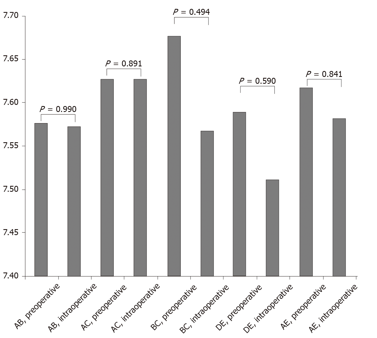
Figure 2 Comparison of preoperative computer tomography measurement data and intraoperative measurement data.
A: The pubic tubercle; B: Intersection of the horizontal line extending from the summit of the inferior edge of the internal oblique and transversus abdominis and the outer edge of the rectus abdominis, C: Intersection of the horizontal line extending from the summit of the inferior edge of the internal oblique and transversus abdominis and the inguinal ligament, D: Intersection of the iliopsoas muscle and the inguinal ligament, and E: Intersection of the iliopsoas muscle and the superior pubic ramus.
- Citation: Wang F, Yang XF. Application of computer tomography-based 3D reconstruction technique in hernia repair surgery. World J Clin Cases 2020; 8(23): 5944-5951
- URL: https://www.wjgnet.com/2307-8960/full/v8/i23/5944.htm
- DOI: https://dx.doi.org/10.12998/wjcc.v8.i23.5944


