Copyright
©The Author(s) 2020.
World J Clin Cases. Nov 26, 2020; 8(22): 5744-5750
Published online Nov 26, 2020. doi: 10.12998/wjcc.v8.i22.5744
Published online Nov 26, 2020. doi: 10.12998/wjcc.v8.i22.5744
Figure 1 Gastroendoscopy findings.
Endoscopic findings showed granular superficial elevated (0-IIa) lesion with a maximum diameter of 10 mm on the anterior wall of the gastric body.
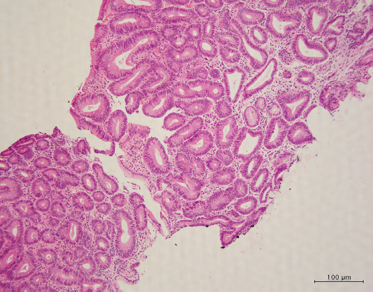
Figure 2 Pathologic examination.
Microscopic examination (hematoxylin and eosin-stain) of biopsy specimen showed many ducts for adenomas, but some of them have structural atypical ducts, suggesting well-differentiated adenocarcinoma.
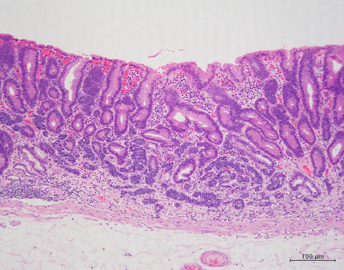
Figure 3 Pathologic examination.
Microscopic examination (hematoxylin and eosin -stain) of resected gastric membrane showed mixture of superficial mucosal adenoma and deep mucosal neuroendocrine tumor.
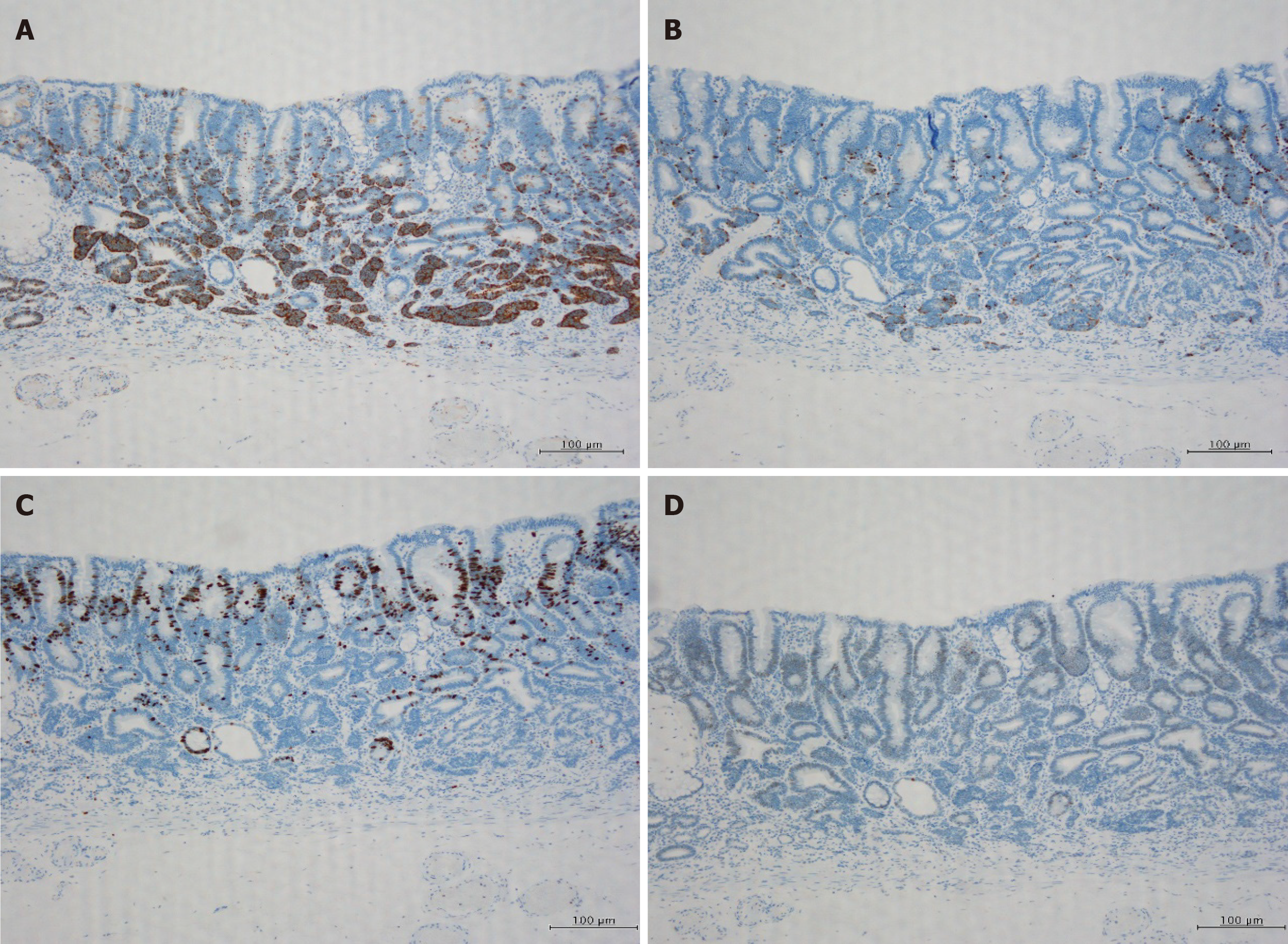
Figure 4 Immunohistochemical staining for synaptophysin, chromogranin, Ki-67, and p-53.
A: Immunohistochemical staining with synaptophysin showed diffuse staining in the cytoplasm of neuroendocrine tumor (NET); B: Immunohistochemical staining with chromogranin slightly stained the NET cytoplasm; C: Immunohistochemical staining with Ki-67 showed a MIB-1 index of 1% or less in the NET region; D: Immunohistochemical staining with p-53 showed almost no staining.
- Citation: Kohno S, Aoki H, Kato M, Ogawa M, Yoshida K. Gastric mixed adenoma-neuroendocrine tumor: A case report. World J Clin Cases 2020; 8(22): 5744-5750
- URL: https://www.wjgnet.com/2307-8960/full/v8/i22/5744.htm
- DOI: https://dx.doi.org/10.12998/wjcc.v8.i22.5744


