Copyright
©The Author(s) 2020.
World J Clin Cases. Nov 26, 2020; 8(22): 5690-5700
Published online Nov 26, 2020. doi: 10.12998/wjcc.v8.i22.5690
Published online Nov 26, 2020. doi: 10.12998/wjcc.v8.i22.5690
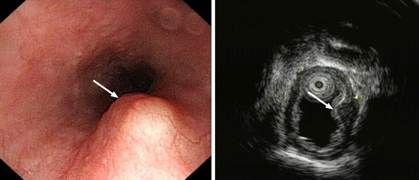
Figure 1 Case 1 Endoscopic view of the submucosal tumor located in the lower thoracic esophagus.
Endoscopic ultrasound revealed a lesion arising from the muscular layer, misdiagnosed as leiomyoma (white arrow).
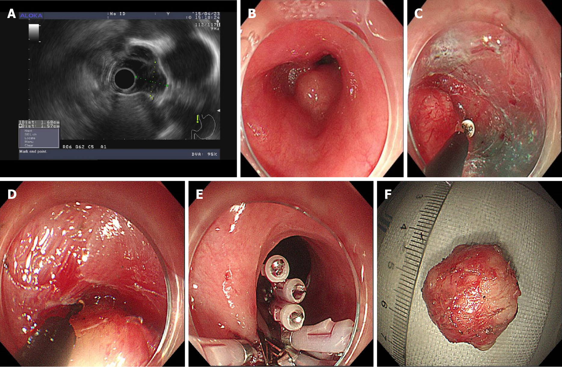
Figure 2 Case 2 Steps of submucosal tunneling endoscopic resection.
A: Endoscopic ultrasound showed the lesion arose from the muscular layer and was misdiagnosed as leiomyoma. The blood flow was not obvious, and the lesion was near the aorta; B: The submucosal tumor located in the middle of the thoracic esophagus; C: A submucosal tunnel was created; D: The tumor was dissected from the muscular layer in the submucosal tunnel; E: Closure of the submucosal tunnel with clips; F: The excised lesion.
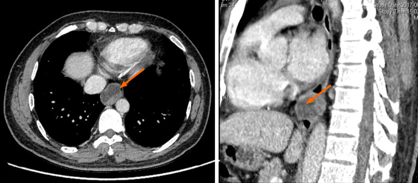
Figure 3 Case 3 Chest computed tomography showed a large mass located in the thoracic esophagus suspected to be a neurogenic tumor or a gastrointestinal stromal tumor (arrow).
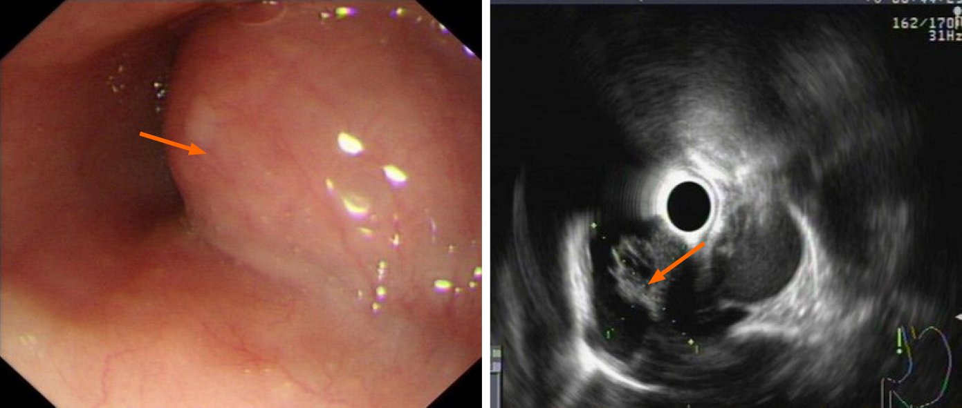
Figure 4 Case 3 Endoscopy revealed a 28 mm × 22 mm submucosal lesion.
Endoscopic ultrasound showed the lesion was derived from the muscular layer with cystic changes and was misdiagnosed as a cystic solid tumor.
Figure 5 Endoscopic submucosal excision performed.
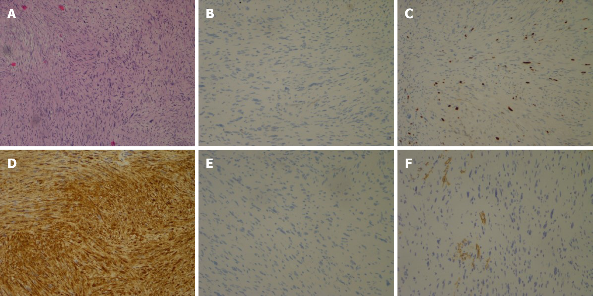
A: Histopathological examination of the tumor in case 1 showed spindle-shaped cells arranged in bundles; B: Immunochemical analysis revealed no staining with CD117; C: Focal positivity with CD34; D: Positive staining with S100; E: DOG-1; F: The mitotic activity was 3 mitosis/50 high-power field on Ki-67 staining.
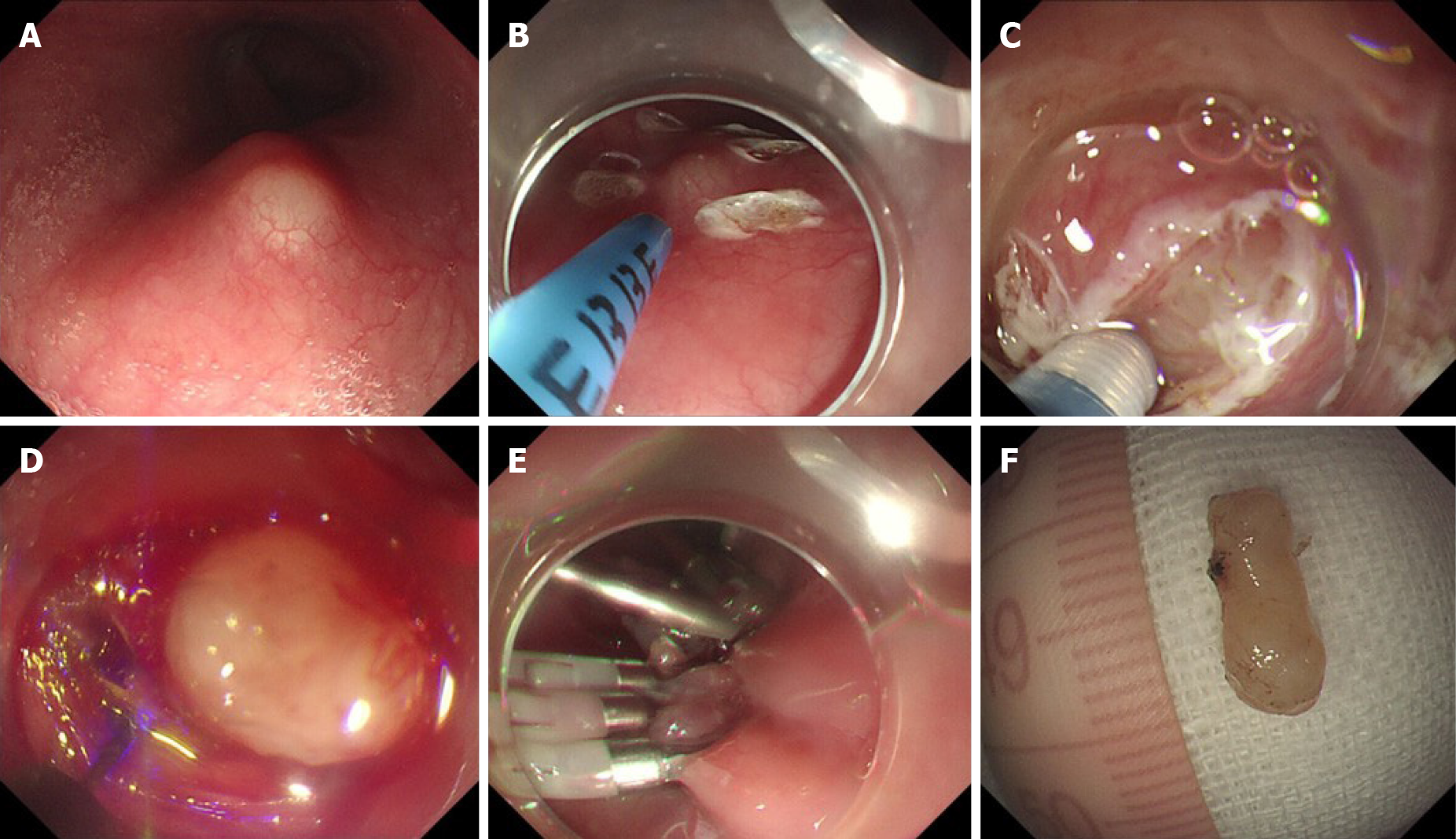
Figure 6 Case 1 Steps of endoscopic submucosal excavation.
A: The submucosal tumor; B: Marking the submucosal tumor with an argon knife; C: Revealing the submucosal tumor; D: Peeling the lesion; E: Closing the mucosal incision site with clips; F: The resected specimen.
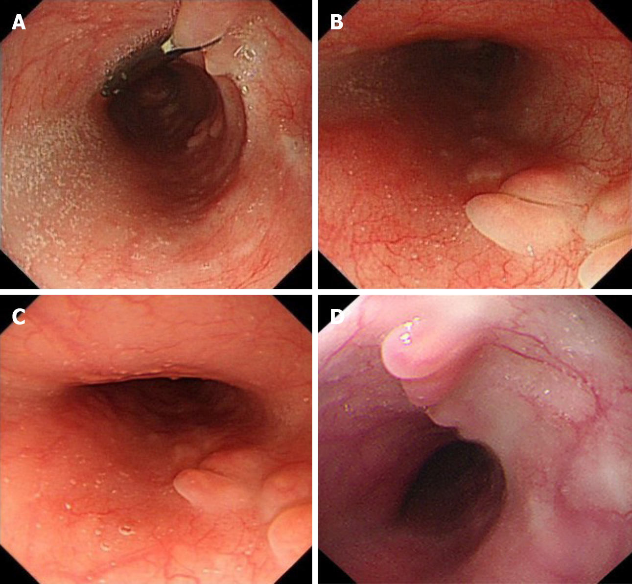
Figure 7 Case 3 Endoscopic follow-up after the endoscopic operation.
A: Residual titanium clip; B: Mucosal scarring was observed at 3 mo. C: Mucosal scarring at 12 mo after the endoscopic operation; D: Mucosal scarring at 24 mo after the endoscopic operation.
- Citation: Li B, Wang X, Zou WL, Yu SX, Chen Y, Xu HW. Endoscopic resection of benign esophageal schwannoma: Three case reports and review of literature. World J Clin Cases 2020; 8(22): 5690-5700
- URL: https://www.wjgnet.com/2307-8960/full/v8/i22/5690.htm
- DOI: https://dx.doi.org/10.12998/wjcc.v8.i22.5690


