Copyright
©The Author(s) 2020.
World J Clin Cases. Nov 26, 2020; 8(22): 5684-5689
Published online Nov 26, 2020. doi: 10.12998/wjcc.v8.i22.5684
Published online Nov 26, 2020. doi: 10.12998/wjcc.v8.i22.5684
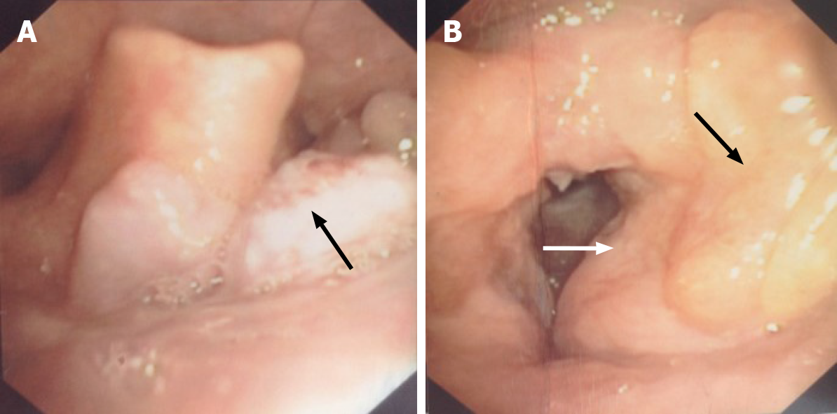
Figure 1 Fiber laryngoscopy findings before surgery.
A: Fiber laryngoscopy showed a neoplasm with a coarse surface (black arrow) near the tongue base; B: Left vocal cord was covered by swelling and thickening ventricular fold (white arrow), and yellow sediment was observed near the left ventricular fold (black arrow). No lesion was found on the right vocal cord.
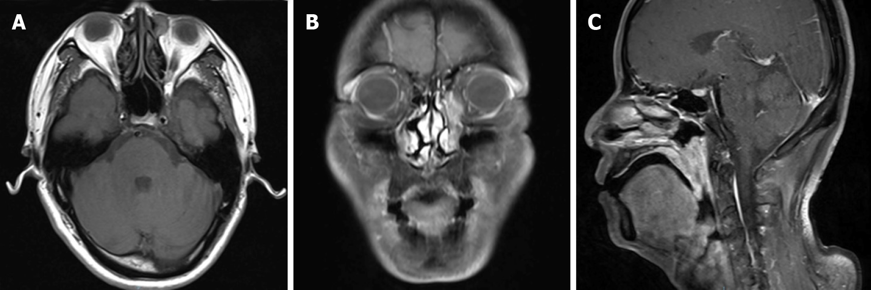
Figure 2 Pre-operative head and neck magnetic resonance imaging.
A: T1 weighted imaging (T1WI) showed a round soft tissue intensity mass within the left lacrimal sac, presenting equal signal in T1WI, and heterogeneous high signal in T2WI, without obvious enhancement when contrasted; B: T1WI with contrast showed an abnormal high signal mass along the left nasolacrimal duct, from the lacrimal sac to the level of inferior turbinate; C: Sagittal T1WI demonstrated extensive soft tissue thickening of the posterior wall of the nasopharynx and the lateral wall of the oropharynx and uvula, with isointensity on T1WI and slightly high signal on T2WI, apparently enhanced when contrasted.
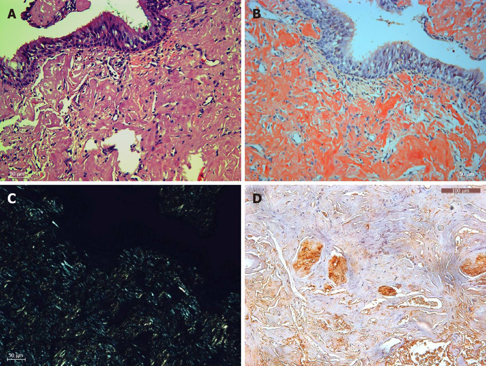
Figure 3 Histopathology (× 200).
A: Hematoxylin and eosin staining of amyloid tissues; B: Congo staining of the same area of amyloid tissues; C: Corresponding area under a polarization microscope; D: Immunohistochemistry showed positive lambda light chain staining. Bar: 100 μm.
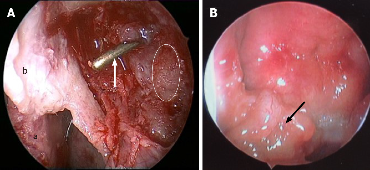
Figure 4 Endoscopic images in surgery.
A: The left lacrimal sac was filled with friable, grey to yellow, glistening, and waxy sediment (white line area), dacryocystorhinostomy was done, and the probe (white arrow) showed opening of the lacrimal sac into the nasal cavity (a: Middle turbinate; b: Nasal mucosa flap); B: An irregular-surfaced mass was noted in the right nasopharynx.
- Citation: Song X, Yang J, Lai Y, Zhou J, Wang J, Sun X, Wang D. Localized amyloidosis affecting the lacrimal sac managed by endoscopic surgery: A case report. World J Clin Cases 2020; 8(22): 5684-5689
- URL: https://www.wjgnet.com/2307-8960/full/v8/i22/5684.htm
- DOI: https://dx.doi.org/10.12998/wjcc.v8.i22.5684


