Copyright
©The Author(s) 2020.
World J Clin Cases. Nov 26, 2020; 8(22): 5639-5644
Published online Nov 26, 2020. doi: 10.12998/wjcc.v8.i22.5639
Published online Nov 26, 2020. doi: 10.12998/wjcc.v8.i22.5639
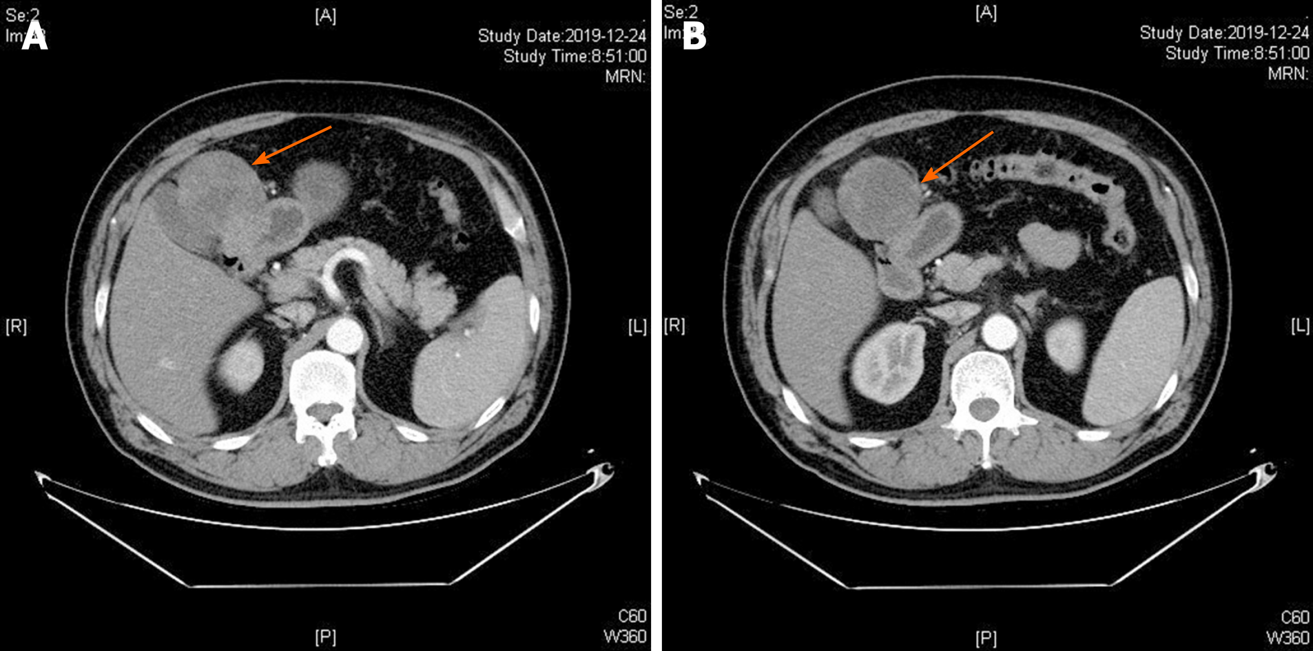
Figure 1 Contrast-enhanced computed tomography axial view revealed a tumor mass with an inhomogeneous enhancement in the arterial phase (A and B).
Figure 2 Microscopic examination showed that the tumor exhibited a multinodular plexiform growth pattern.
These nodules consisted of bland-looking spindle cells admixed with abundant myxoid or fibromyxoid stroma rich in capillary-sized vessels (hematoxylin and eosin, A: × 100; B: × 400).
Figure 3 Immunohistochemical examinations of the tumor (× 400).
Tumor cells showed diffuse cytoplasmic positivity for smooth muscle actin (A) and S-100 (B), but were negative for CD117 (C).
- Citation: Pei JY, Tan B, Liu P, Cao GH, Wang ZS, Qu LL. Gastric plexiform fibromyxoma: A case report. World J Clin Cases 2020; 8(22): 5639-5644
- URL: https://www.wjgnet.com/2307-8960/full/v8/i22/5639.htm
- DOI: https://dx.doi.org/10.12998/wjcc.v8.i22.5639


