Copyright
©The Author(s) 2020.
World J Clin Cases. Aug 26, 2020; 8(16): 3583-3590
Published online Aug 26, 2020. doi: 10.12998/wjcc.v8.i16.3583
Published online Aug 26, 2020. doi: 10.12998/wjcc.v8.i16.3583
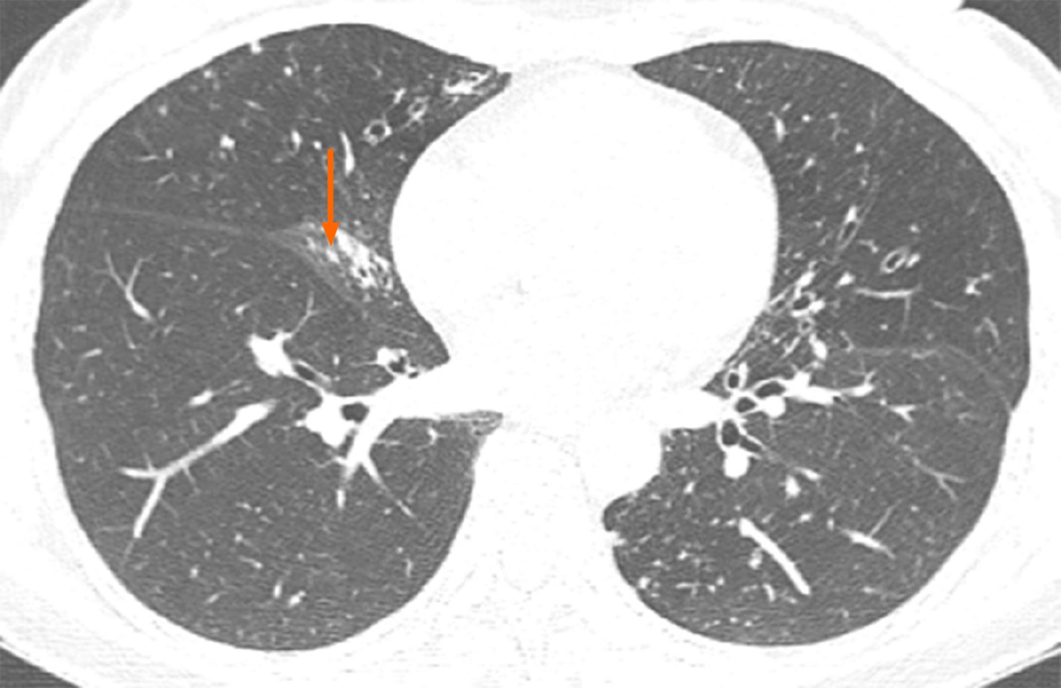
Figure 1 In November 2017, computed tomography showed atrophy in the middle lobe of her right lung as well as bronchiectasis in the left lung and the middle lobe of the right lung accompanied by infections.
Numerous nodules with a diameter of 0.3 to 0.5 cm were identified in the middle lobe of the right lung (arrow).
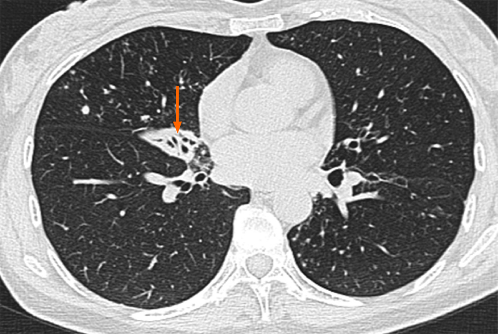
Figure 2 Another computed tomography image obtained in March 2018 revealed that the bronchiectasis in the middle lobe of her right lung was accompanied by atelectasis (arrow), which was more observable than the atrophy in the same location in the previous computed tomography image.
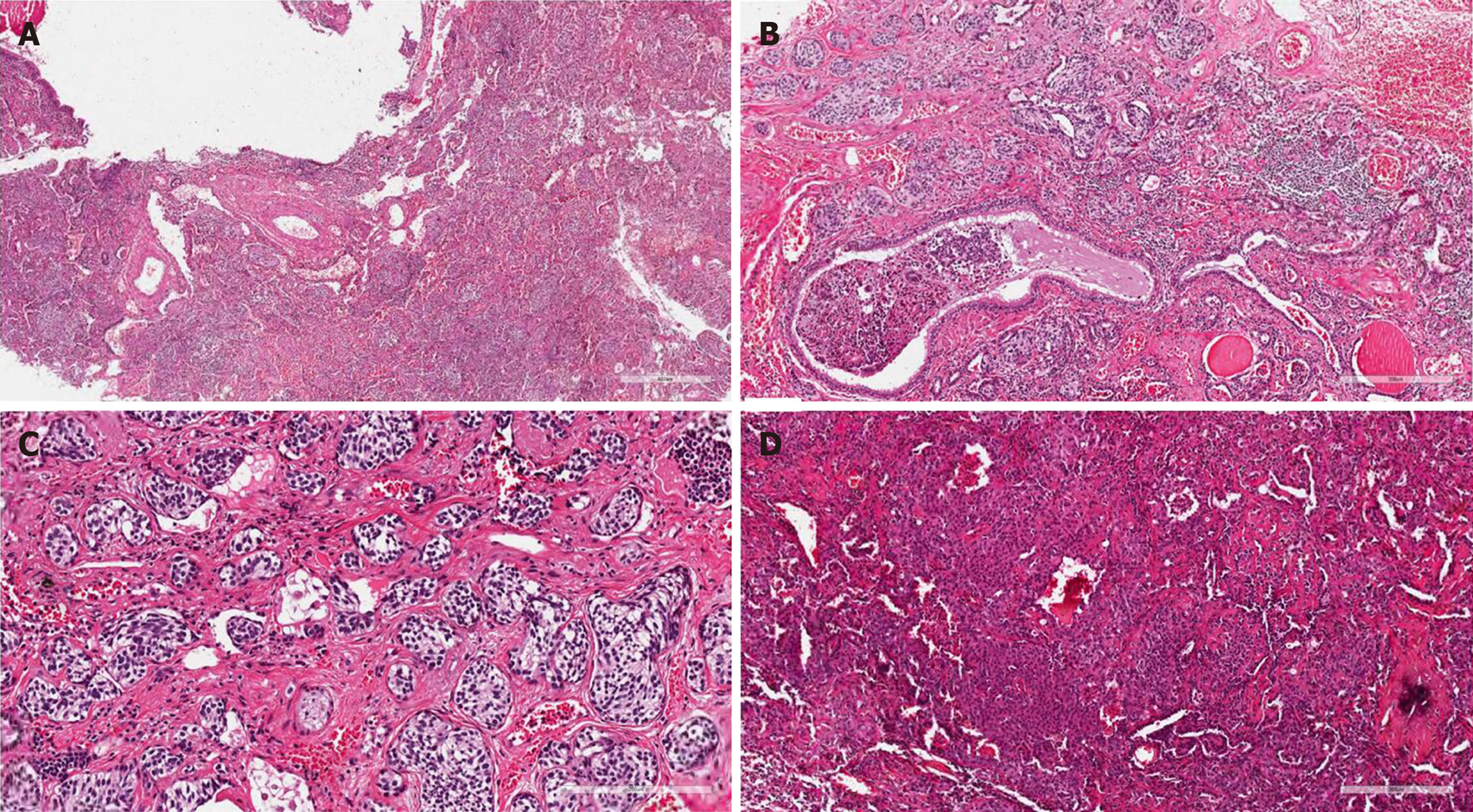
Figure 3 Pathological findings.
A: Microscopic examination showed inflammatory cell infiltration with lymphocytes, neutrophils, and histiocytes in alveoli. Alveolar septa broke and merged, focal bronchiectasis was observed, and cartilage in the bronchus was damaged [(hematoxylin and eosin staining (HE staining), × 40]; B: Multifocal neuroendocrine-like cell nest hyperplasia was found in the surroundings of the expanded bronchus (single focal diameter < 0.5 cm; HE staining, × 100); C: The nuclei of hyperplasia cells are polygonal or short fusiform with a uniform size. The nuclear chromatin was delicate and in fine particles. The nucleoli were unclear; no mitotic figures or necrosis was detected (HE staining, × 200); D: HE staining (× 100) showed the papillary arrangement of tumor cells and a sample of vascular cavity.
- Citation: Han XY, Wang YY, Wei HQ, Yang GZ, Wang J, Jia YZ, Ao WQ. Multifocal neuroendocrine cell hyperplasia accompanied by tumorlet formation and pulmonary sclerosing pneumocytoma: A case report. World J Clin Cases 2020; 8(16): 3583-3590
- URL: https://www.wjgnet.com/2307-8960/full/v8/i16/3583.htm
- DOI: https://dx.doi.org/10.12998/wjcc.v8.i16.3583


