Copyright
©The Author(s) 2020.
World J Clin Cases. Jul 26, 2020; 8(14): 3064-3073
Published online Jul 26, 2020. doi: 10.12998/wjcc.v8.i14.3064
Published online Jul 26, 2020. doi: 10.12998/wjcc.v8.i14.3064
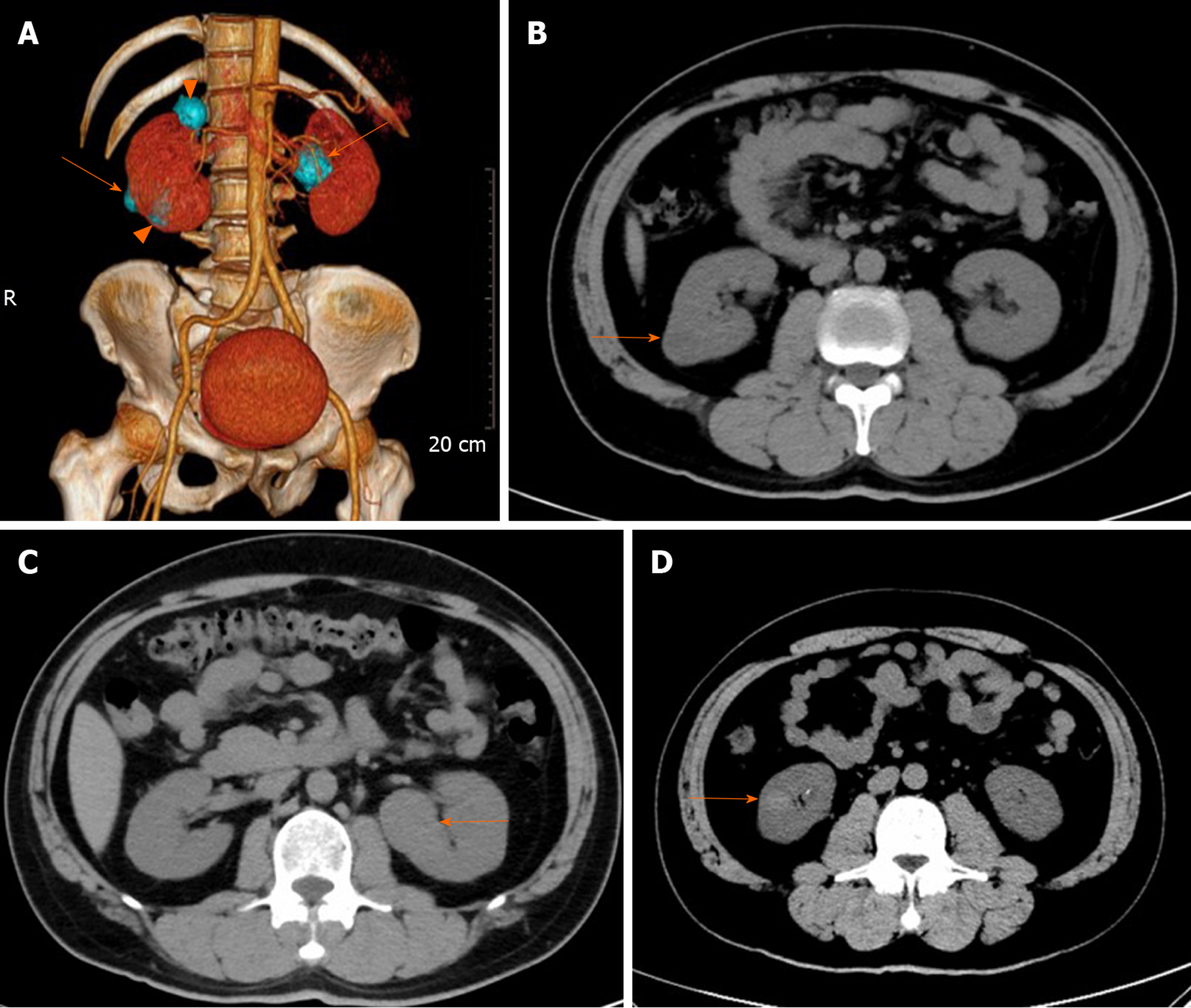
Figure 1 Computed tomography imaging of the patient.
A: Volume representation shows bilateral multiple renal tumors (4 masses); B: Pre-operative axial computed tomography (CT) imaging sections showing a 1.9 cm × 1.9 cm × 2.0 cm tumor arising from the right kidney, exophytic, heterogeneous hypodense, without calcification and hemorrhage, proven to be chromophobe renal cell carcinoma (orange arrow); C: Axial CT imaging sections showing a 3.3 cm × 3.2 cm × 2.9 cm tumor arising from the left kidney, exophytic, homogeneous isodense, without calcification and hemorrhage, proven to be a chromophobe renal cell carcinoma (orange arrow); and D: Axial CT imaging sections showing a 1.4 cm × 1.3 cm × 1.1 cm tumor arising from the right kidney, homogeneous hyperdense, without calcification and hemorrhage, proven to be a clear cell renal cell carcinoma (orange arrow).
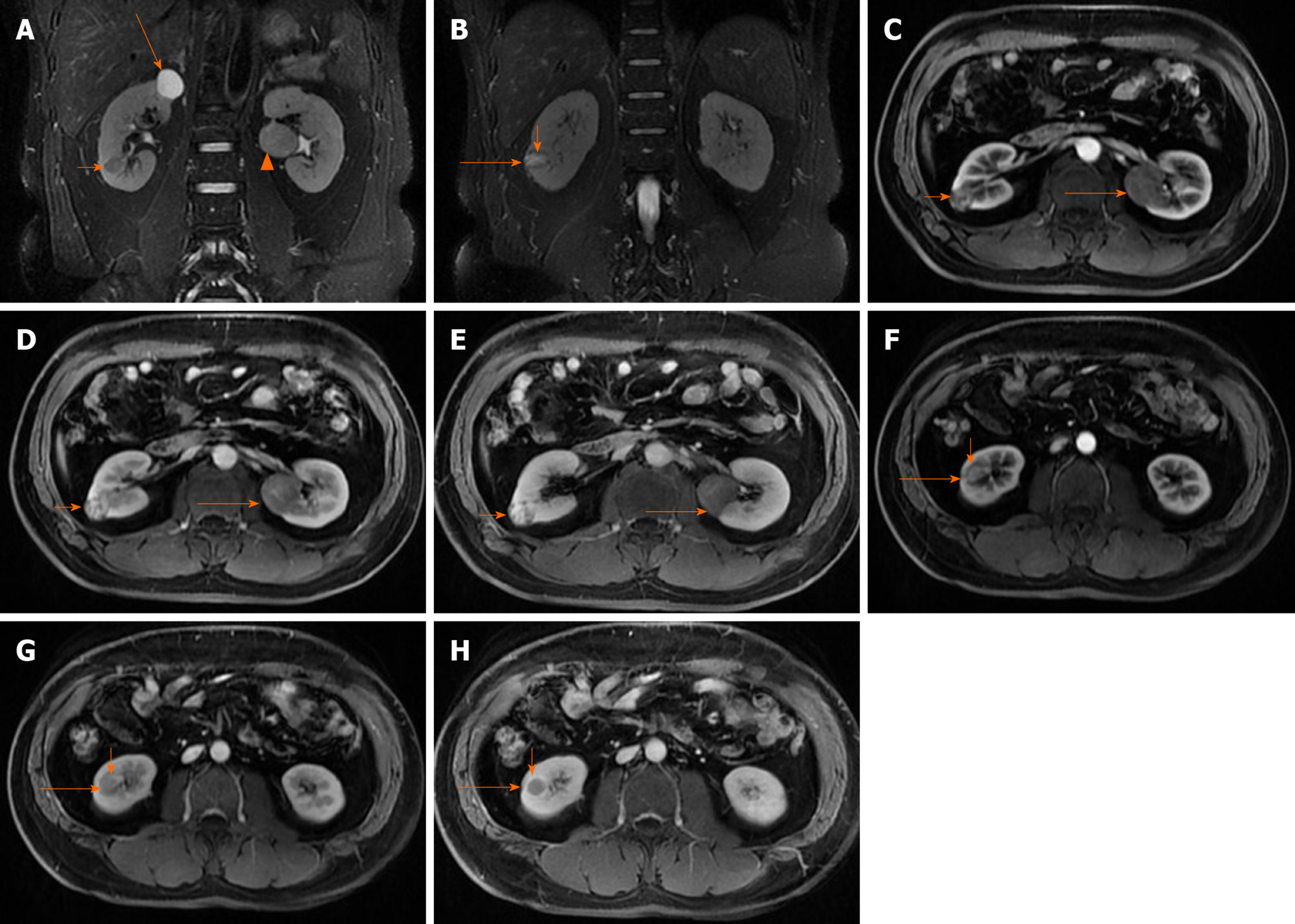
Figure 2 Magnetic resonance imaging of the patient.
A: Pre-operative coronal fat-saturated series T2-weighted image of the patient demonstrating a cystic lesion at the upper pole of the right kidney showing strongly signal intensity (long arrow), and a homogeneous low signal intensity, solid mass at right kidney proven to be clear cell renal cell carcinoma (short arrow), and a solid mass, intermediate signal intensity in relation to the adjacent cortex, with a distinct hyposignal pseudocapsule at left kidney proven to be chromophobe renal cell carcinoma (CHRCC) (arrowhead); B: A heterogeneous hypersignal intensity (long arrow) with local cystic, with a distinct hyposignal pseudocapsule (short arrow) on right kidney, proven to be an oncocytic variant of CHRCC; C-E: Axial Contrast-enhanced magnetic resonance image shows heterogeneous signal intensity mass in the right kidney, heterogeneous delayed enhancement, multiple small cysts without enhancement, proven to be an oncocytic variant of CHRCC (short arrow), and a solid mass in the middle of the left kidney, tend to present with progressive uptake in the contrast-enhanced magnetic resonance imaging (MRI), diagnosed as CHRCC (long arrow); F-H: Axial contrast-enhanced MRI shows a solid mass in the right kidney (long arrow), tends to present “fast in and fast out” pattern in contrast-enhanced MRI, and pseudocapsule (short arrow) delayed enhancement, diagnosed as clear cell renal cell carcinoma.
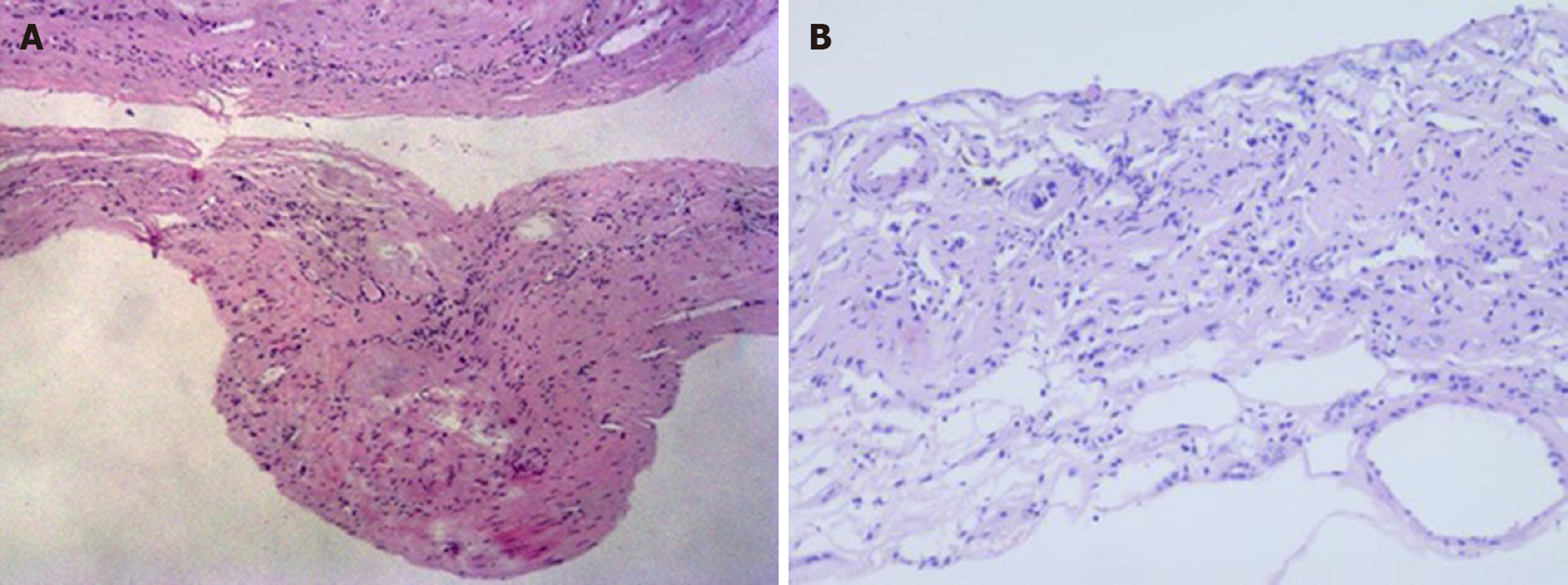
Figure 3 A 50-year-old male with a pathologically proven cyst in the right kidney.
A: H&E staining showed fibromuscular tissue capsule walls without epithelial lining, in which atrophic tubular structures (× 40); B: Simple cyst (× 100).
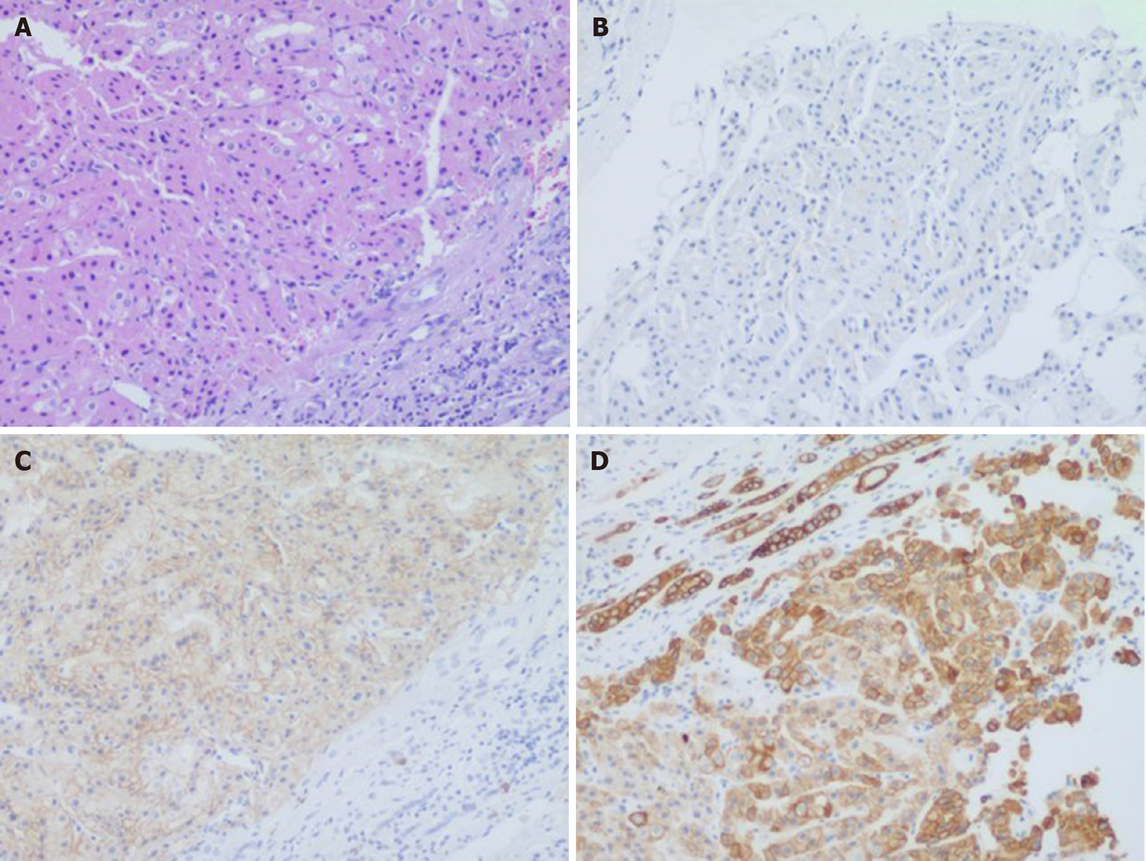
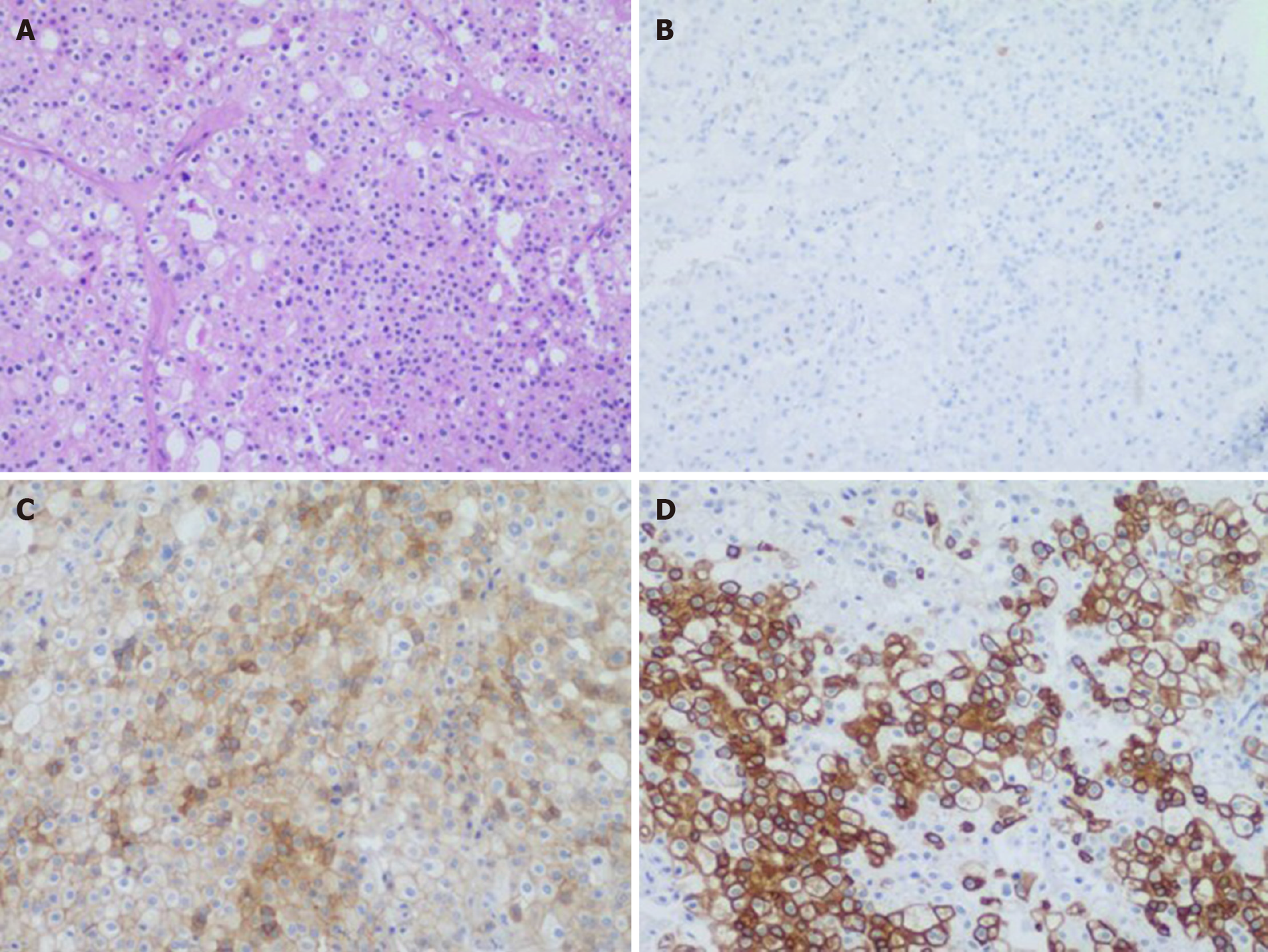
Figure 4 A 50-year-old male with a pathologically proven chromophobe renal cell carcinoma in the right kidney (eosinophilic type).
A: Hematoxylin and eosin staining showing tubular or acinar structures, eosinophilic to pale cytoplasm with accentuated cell borders, and “raisinoid” nuclear membranes (100 ×); B: Immunohistochemical staining showing negative expression of carbonic anhydrase 9 (100 ×); C: Immunohistochemical staining showing cluster of differentiation 117 was moderately diffusely positive in the cytoplasm/membrane (100 ×); D: Immunohistochemical staining showing cytokeratin 7 (D) was strongly diffusely positive in the cytoplasm/membrane (100 ×).
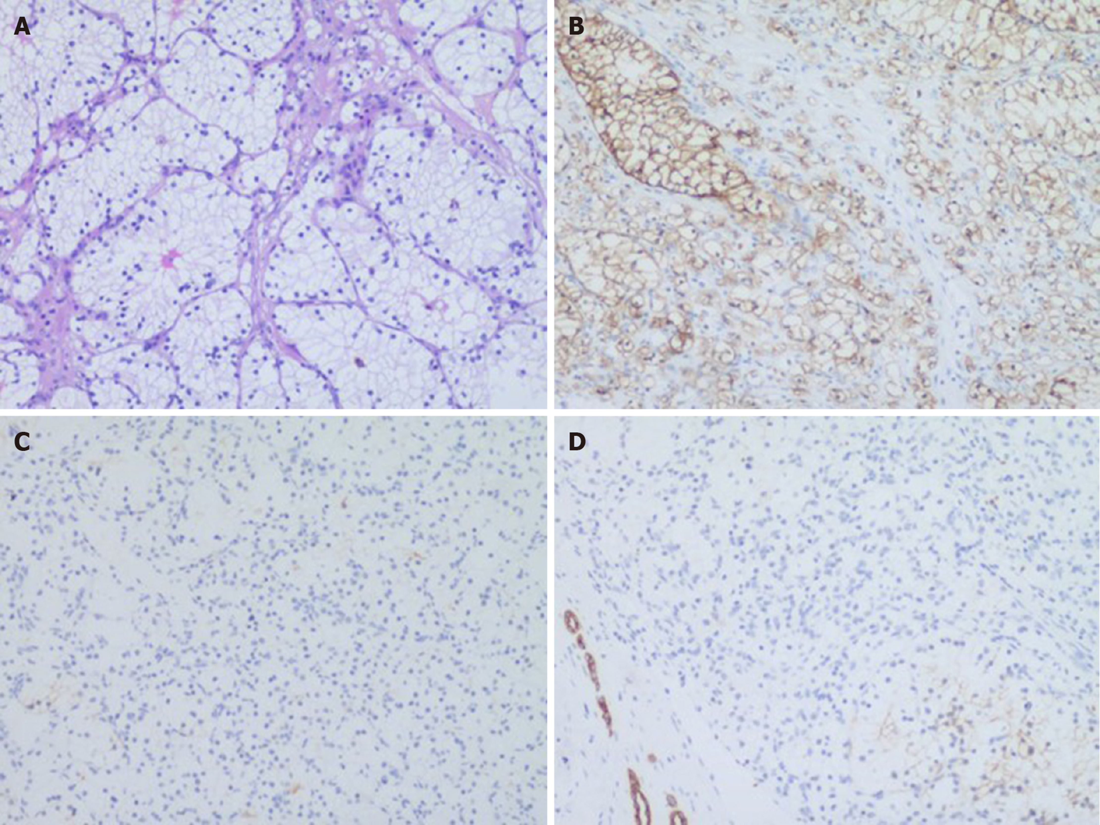
Figure 5 A 50-year-old male with pathologically proven clear cell renal cell carcinoma in the right kidney.
A: Hematoxylin and eosin staining showing nesting and tubular pattern of growth with eosinophilic cytoplasm in delicate vascular network, clear cytoplasm (100 ×); B: Immunohistochemical staining, carbonic anhydrase 9 was strongly diffusely positive in the cytoplasm/membrane (100 ×); C: Immunohistochemical staining showing negative expression of cluster of differentiation 117 (× 100); D: Immunohistochemical staining showing negative expression of cytokeratin 7 (100 ×).
Figure 6 A 50-year-old male with pathologically proven chromophobe renal cell carcinoma in the left kidney.
A: Hematoxylin and eosin staining showing tumor cells were arranged in solid nests and gland pattern with thick-walled blood vessels, clear cell membrane, reticulated cytoplasm, and “raisinoid” nuclear membranes (100 ×); B: Immunohistochemical staining showing negative expression of carbonic anhydrase 9 (100 ×); C: Cluster of differentiation 117 was moderately diffusely positive in the cytoplasm/membrane (100 ×); D: Cytokeratin 7 was strongly diffusely positive in the cytoplasm/membrane (100 ×).
- Citation: Yang F, Zhao ZC, Hu AJ, Sun PF, Zhang B, Yu MC, Wang J. Synchronous sporadic bilateral multiple chromophobe renal cell carcinoma accompanied by a clear cell carcinoma and a cyst: A case report. World J Clin Cases 2020; 8(14): 3064-3073
- URL: https://www.wjgnet.com/2307-8960/full/v8/i14/3064.htm
- DOI: https://dx.doi.org/10.12998/wjcc.v8.i14.3064


