Copyright
©The Author(s) 2020.
World J Clin Cases. Jun 26, 2020; 8(12): 2662-2666
Published online Jun 26, 2020. doi: 10.12998/wjcc.v8.i12.2662
Published online Jun 26, 2020. doi: 10.12998/wjcc.v8.i12.2662
Figure 1 Chest high-resolution computed tomography.
A: Reduced lucency of both lungs, patchy density and ground-glass opacity were seen in multiple lobes with fuzzy boundaries; B: Diffuse cystic lesions in both lungs and patchy opacity with fuzzy boundaries in multiple lobes; C: Diffuse ground-glass opacity was observed in both lungs with fuzzy boundaries.
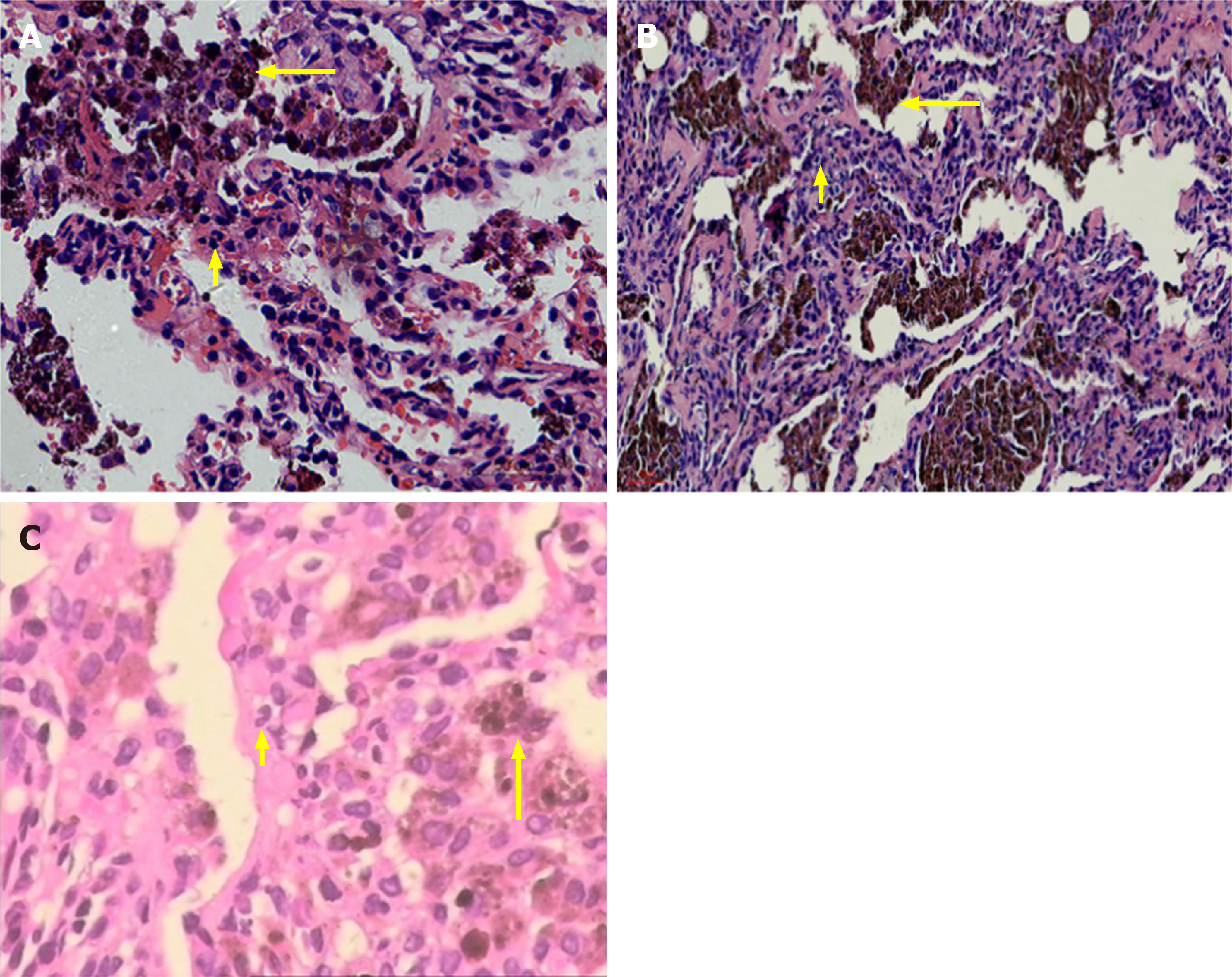
Figure 2 Hematoxylin and eosin staining.
A: Aggregation of hemosiderin-laden macrophage cells (HLMs) within the alveolar compartment (long arrow), alveolar interval are mild fibrous thickening and neutrophil infiltrate (short arrow) (400 ×); B: Acute extravasation of fibrin and red blood cells, HLM deposits in the alveolar cavities (long arrow) and neutrophil infiltrates (short arrow) (400 ×); C: Partial lung tissue fibrosis and HLM deposits were observed in the alveolar cavities (long arrow) and neutrophil infiltrates (short arrow) (400 ×).
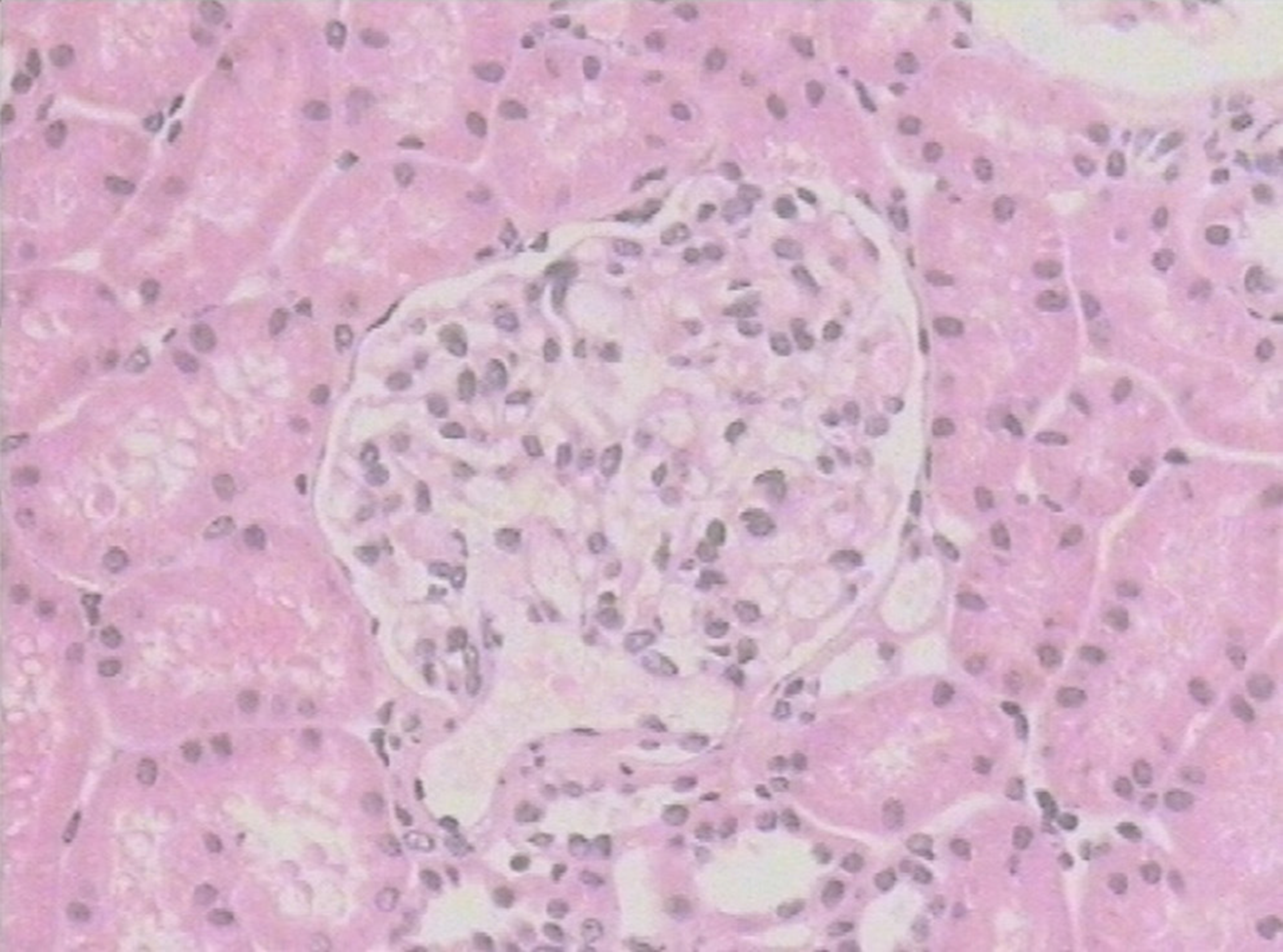
Figure 3 Hematoxylin and eosin staining.
Twenty nonsclerotic glomeruli were identified by light microscopy, which mainly presented as mesenteric proliferative changes. No glomeruli contained fibrous crescents and no proliferative changes were observed in the endocapillary or extracapillary areas (400 ×).
- Citation: Xie J, Zhao YY, Liu J, Nong GM. Diffuse alveolar hemorrhage with histopathologic manifestations of pulmonary capillaritis: Three case reports. World J Clin Cases 2020; 8(12): 2662-2666
- URL: https://www.wjgnet.com/2307-8960/full/v8/i12/2662.htm
- DOI: https://dx.doi.org/10.12998/wjcc.v8.i12.2662


